4GH7

Crystal structure of Anticalin N7A in complex with oncofetal fibronectin fragment Fn7B8


Experimental Data Snapshot

  • Method: X-RAY DIFFRACTION
  • Resolution: 2.60 Å
  • R-Value Free: 0.259 
  • R-Value Work: 0.218 
  • R-Value Observed: 0.220 

wwPDB Validation   3D Report Full Report


This is version 1.3 of the entry. See complete history


Literature

Combinatorial design of an Anticalin directed against the extra-domain b for the specific targeting of oncofetal fibronectin

Gebauer, M.Schiefner, A.Matschiner, G.Skerra, A.

(2013) J Mol Biol 425: 780-802

  • DOI: https://doi.org/10.1016/j.jmb.2012.12.004
  • Primary Citation of Related Structures:  
    4GH7

  • PubMed Abstract: 

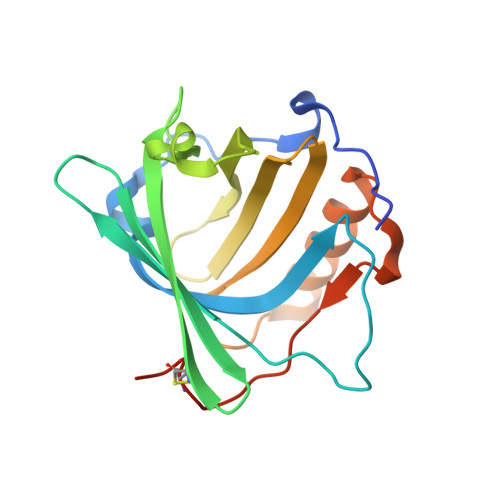
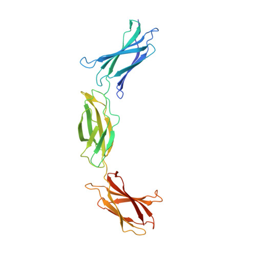
    The oncofetal isoform of the extracellular matrix protein fibronectin (Fn), which carries the extra-domain B (ED-B) and is exclusively expressed in neovasculature, has gained interest for tumor diagnosis and therapy using engineered antibody fragments. We have employed the human lipocalin 2 (Lcn2) as a small and robust non-immunoglobulin scaffold to select ED-B-specific Anticalins from a new advanced random library using bacterial phage display and ELISA screening against appropriately engineered Fn fragments. As a result, we have isolated and biochemically characterized four different Anticalins that all show low nanomolar affinities for ED-B, right in the range between the monomeric and dimeric forms of the single-chain variable antibody fragment L19 that has been widely applied in this area before. All Anticalins can be readily expressed in Escherichia coli as soluble and strictly monomeric proteins, and they show specific staining of ED-B-positive tumor cells in immunofluorescence microscopy while BIAcore affinity analyses indicate recognition of distinct ED-B epitopes. The crystal structure for one Anticalin, N7A, in complex with the Fn7B8 fragment, was solved at 2.6Å resolution and reveals binding to the gfcc' sheet and cc' loop on ED-B. This is the second example of a protein-specific Lcn2-based Anticalin, which illustrates the remarkable plasticity of the calyx-like ligand pocket of lipocalins with their four structurally hypervariable loops supported by a highly conserved β-barrel. The ED-B-specific Anticalins resulting from this study should provide useful reagents in research and biomedical drug development, both for in vivo imaging and for directed cancer therapy.


  • Organizational Affiliation

    Munich Center for Integrated Protein Science (CIPS-M) and Lehrstuhl für Biologische Chemie, Technische Universität München, 85350 Freising-Weihenstephan, Germany.


Macromolecules
Find similar proteins by:  (by identity cutoff)  |  3D Structure
Entity ID: 1
MoleculeChains Sequence LengthOrganismDetailsImage
Neutrophil gelatinase-associated lipocalin
A, C
188Homo sapiensMutation(s): 21 
Gene Names: HNLLCN2NGAL
UniProt & NIH Common Fund Data Resources
Find proteins for P80188 (Homo sapiens)
Explore P80188 
Go to UniProtKB:  P80188
PHAROS:  P80188
GTEx:  ENSG00000148346 
Entity Groups  
Sequence Clusters30% Identity50% Identity70% Identity90% Identity95% Identity100% Identity
UniProt GroupP80188
Sequence Annotations
Expand
  • Reference Sequence
Find similar proteins by:  (by identity cutoff)  |  3D Structure
Entity ID: 2
MoleculeChains Sequence LengthOrganismDetailsImage
Fibronectin
B, D
285Homo sapiensMutation(s): 0 
Gene Names: FN1
UniProt & NIH Common Fund Data Resources
Find proteins for P02751 (Homo sapiens)
Explore P02751 
Go to UniProtKB:  P02751
PHAROS:  P02751
GTEx:  ENSG00000115414 
Entity Groups  
Sequence Clusters30% Identity50% Identity70% Identity90% Identity95% Identity100% Identity
UniProt GroupP02751
Sequence Annotations
Expand
  • Reference Sequence
Experimental Data & Validation

Experimental Data

  • Method: X-RAY DIFFRACTION
  • Resolution: 2.60 Å
  • R-Value Free: 0.259 
  • R-Value Work: 0.218 
  • R-Value Observed: 0.220 
  • Space Group: P 43 21 2
Unit Cell:
Length ( Å )Angle ( ˚ )
a = 107.126α = 90
b = 107.126β = 90
c = 233.156γ = 90
Software Package:
Software NamePurpose
XSCALEdata scaling
REFMACrefinement
PDB_EXTRACTdata extraction
MAR345data collection
XDSdata reduction
PHASERphasing

Structure Validation

View Full Validation Report


Entry History 

Deposition Data

Revision History  (Full details and data files)

  • Version 1.0: 2012-12-26
    Type: Initial release
  • Version 1.1: 2013-04-10
    Changes: Database references
  • Version 1.2: 2017-11-15
    Changes: Refinement description
  • Version 1.3: 2023-11-08
    Changes: Data collection, Database references, Refinement description