4GW9

Structure of a bacteriophytochrome and light-stimulated protomer swapping with a gene repressor


Experimental Data Snapshot

  • Method: X-RAY DIFFRACTION
  • Resolution: 2.90 Å
  • R-Value Free: 0.243 
  • R-Value Work: 0.200 
  • R-Value Observed: 0.202 

wwPDB Validation   3D Report Full Report


Ligand Structure Quality Assessment 


This is version 1.0 of the entry. See complete history


Literature

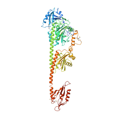
Structure of a bacteriophytochrome and light-stimulated protomer swapping with a gene repressor.

Bellini, D.Papiz, M.Z.

(2012) Structure 20: 1436-1446

  • DOI: https://doi.org/10.1016/j.str.2012.06.002
  • Primary Citation of Related Structures:  
    4GW9

  • PubMed Abstract: 

    Phytochromes are photoreceptors in phototropic organisms that respond to light conditions by changing interactions between a response regulator and DNA. Bacterial phytochromes (BphPs) comprise an input photosensory core domain (PCD) and an output transducing domain (OTD). We report the structure of a BphP containing both PCD and the majority of its OTD, and demonstrate interaction with its cognate repressor. The OTD of RpBphP1, from Rhodopseudomonas palustris, is composed of a PAS/PAC domain and, to our knowledge, a hitherto unrecognized two-helix output sensor (HOS) domain. Unlike canonical BphPs, it does not transmit phosphorelay signals but forms a complex with the transcriptional repressor RpPpsR2 on photoconversion with far-red light. We show that HOS is essential for complex formation and that the anti-parallel dimer geometry is crucial in achieving HOS domain activation and protomer swapping under the control of light. These results provide insights into the steps taken by a two-component signaling system.


  • Organizational Affiliation

    Institute of Integrative Biology, Biosciences Building, University of Liverpool, Crown Street, Liverpool L69 7ZB, UK.


Macromolecules
Find similar proteins by:  (by identity cutoff)  |  3D Structure
Entity ID: 1
MoleculeChains Sequence LengthOrganismDetailsImage
bacteriophytochrome
A, B, C, D
655Rhodopseudomonas palustris CGA009Mutation(s): 0 
Gene Names: RPA1537
EC: 2.7.13.3
UniProt
Find proteins for B3Q7C0 (Rhodopseudomonas palustris (strain TIE-1))
Explore B3Q7C0 
Go to UniProtKB:  B3Q7C0
Entity Groups  
Sequence Clusters30% Identity50% Identity70% Identity90% Identity95% Identity100% Identity
UniProt GroupB3Q7C0
Sequence Annotations
Expand
  • Reference Sequence
Experimental Data & Validation

Experimental Data

  • Method: X-RAY DIFFRACTION
  • Resolution: 2.90 Å
  • R-Value Free: 0.243 
  • R-Value Work: 0.200 
  • R-Value Observed: 0.202 
  • Space Group: P 1 21 1
Unit Cell:
Length ( Å )Angle ( ˚ )
a = 102.94α = 90
b = 146.87β = 101.17
c = 139.55γ = 90
Software Package:
Software NamePurpose
GDAdata collection
SHARPphasing
REFMACrefinement
xia2data reduction
XDSdata reduction
SCALAdata scaling

Structure Validation

View Full Validation Report


Ligand Structure Quality Assessment 


Entry History 

Deposition Data

Revision History  (Full details and data files)

  • Version 1.0: 2012-09-26
    Type: Initial release