4HSA

Structure of interleukin 17a in complex with il17ra receptor


Experimental Data Snapshot

  • Method: X-RAY DIFFRACTION
  • Resolution: 3.15 Å
  • R-Value Free: 0.232 
  • R-Value Work: 0.187 
  • R-Value Observed: 0.189 

wwPDB Validation   3D Report Full Report


Ligand Structure Quality Assessment 


This is version 2.0 of the entry. See complete history


Literature

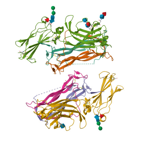
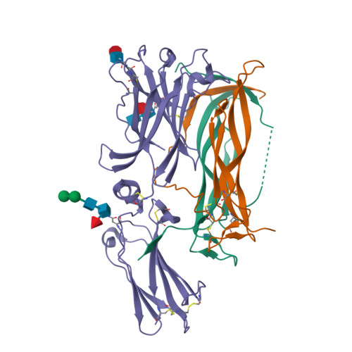
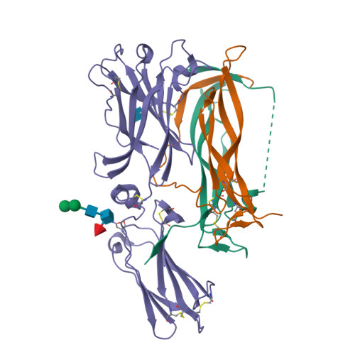
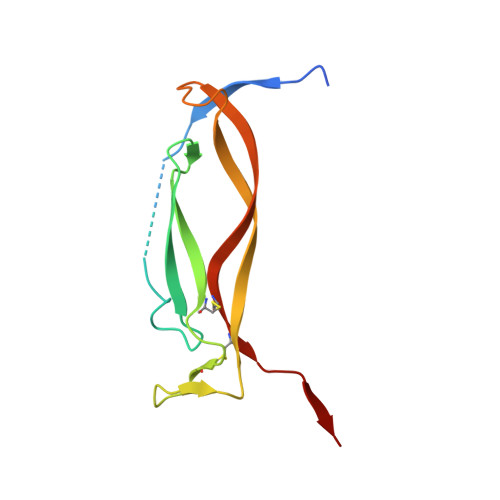
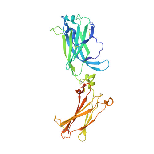
Crystal structures of interleukin 17A and its complex with IL-17 receptor A.

Liu, S.Song, X.Chrunyk, B.A.Shanker, S.Hoth, L.R.Marr, E.S.Griffor, M.C.

(2013) Nat Commun 4: 1888-1888

  • DOI: https://doi.org/10.1038/ncomms2880
  • Primary Citation of Related Structures:  
    4HR9, 4HSA

  • PubMed Abstract: 

    The constituent polypeptides of the interleukin-17 family form six different homodimeric cytokines (IL-17A-F) and the heterodimeric IL-17A/F. Their interactions with IL-17 receptors A-E (IL-17RA-E) mediate host defenses while also contributing to inflammatory and autoimmune responses. IL-17A and IL-17F both preferentially engage a receptor complex containing one molecule of IL-17RA and one molecule of IL-17RC. More generally, IL-17RA appears to be a shared receptor that pairs with other members of its family to allow signaling of different IL-17 cytokines. Here we report crystal structures of homodimeric IL-17A and its complex with IL-17RA. Binding to IL-17RA at one side of the IL-17A molecule induces a conformational change in the second, symmetry-related receptor site of IL-17A. This change favors, and is sufficient to account for, the selection of a different receptor polypeptide to complete the cytokine-receptor complex. The structural results are supported by biophysical studies with IL-17A variants produced by site-directed mutagenesis.


  • Organizational Affiliation

    Structural Biology and Biophysics Group, Pfizer Groton Laboratories, Eastern Point Road, Groton, Connecticut 06340, USA. shenping.liu@pfizer.com


Macromolecules
Find similar proteins by:  (by identity cutoff)  |  3D Structure
Entity ID: 1
MoleculeChains Sequence LengthOrganismDetailsImage
Interleukin-17A
A, B, D, E
122Homo sapiensMutation(s): 2 
Gene Names: IL17ACTLA8IL17
UniProt & NIH Common Fund Data Resources
Find proteins for Q16552 (Homo sapiens)
Explore Q16552 
Go to UniProtKB:  Q16552
PHAROS:  Q16552
GTEx:  ENSG00000112115 
Entity Groups  
Sequence Clusters30% Identity50% Identity70% Identity90% Identity95% Identity100% Identity
UniProt GroupQ16552
Sequence Annotations
Expand
  • Reference Sequence
Find similar proteins by:  (by identity cutoff)  |  3D Structure
Entity ID: 2
MoleculeChains Sequence LengthOrganismDetailsImage
Interleukin-17 receptor A
C, F
301Homo sapiensMutation(s): 2 
Gene Names: IL17RAIL17R
UniProt & NIH Common Fund Data Resources
Find proteins for Q96F46 (Homo sapiens)
Explore Q96F46 
Go to UniProtKB:  Q96F46
PHAROS:  Q96F46
GTEx:  ENSG00000177663 
Entity Groups  
Sequence Clusters30% Identity50% Identity70% Identity90% Identity95% Identity100% Identity
UniProt GroupQ96F46
Sequence Annotations
Expand
  • Reference Sequence
Oligosaccharides

Help

Entity ID: 3
MoleculeChains Length2D Diagram Glycosylation3D Interactions
alpha-D-mannopyranose-(1-3)-alpha-D-mannopyranose-(1-4)-2-acetamido-2-deoxy-beta-D-glucopyranose-(1-4)-[alpha-L-fucopyranose-(1-6)]2-acetamido-2-deoxy-beta-D-glucopyranose
G, J
5N-Glycosylation
Glycosylation Resources
GlyTouCan:  G26032DN
GlyCosmos:  G26032DN
GlyGen:  G26032DN
Entity ID: 4
MoleculeChains Length2D Diagram Glycosylation3D Interactions
2-acetamido-2-deoxy-beta-D-glucopyranose-(1-4)-[alpha-L-fucopyranose-(1-6)]2-acetamido-2-deoxy-beta-D-glucopyranose
H
3N-Glycosylation
Glycosylation Resources
GlyTouCan:  G21290RB
GlyCosmos:  G21290RB
GlyGen:  G21290RB
Entity ID: 5
MoleculeChains Length2D Diagram Glycosylation3D Interactions
alpha-L-fucopyranose-(1-6)-2-acetamido-2-deoxy-beta-D-glucopyranose
I
2N-Glycosylation
Glycosylation Resources
GlyTouCan:  G86851RC
GlyCosmos:  G86851RC
GlyGen:  G86851RC
Small Molecules
Ligands 1 Unique
IDChains Name / Formula / InChI Key2D Diagram3D Interactions
NAG
Query on NAG

Download Ideal Coordinates CCD File 
K [auth F]2-acetamido-2-deoxy-beta-D-glucopyranose
C8 H15 N O6
OVRNDRQMDRJTHS-FMDGEEDCSA-N
Experimental Data & Validation

Experimental Data

  • Method: X-RAY DIFFRACTION
  • Resolution: 3.15 Å
  • R-Value Free: 0.232 
  • R-Value Work: 0.187 
  • R-Value Observed: 0.189 
  • Space Group: P 31 2 1
Unit Cell:
Length ( Å )Angle ( ˚ )
a = 138.71α = 90
b = 138.71β = 90
c = 179.09γ = 120
Software Package:
Software NamePurpose
BUSTERrefinement

Structure Validation

View Full Validation Report


Ligand Structure Quality Assessment 


Entry History 

Deposition Data

  • Released Date: 2013-05-22 
  • Deposition Author(s): Liu, S.

Revision History  (Full details and data files)

  • Version 1.0: 2013-05-22
    Type: Initial release
  • Version 1.1: 2013-06-05
    Changes: Database references
  • Version 1.2: 2013-06-19
    Changes: Database references
  • Version 2.0: 2020-07-29
    Type: Remediation
    Reason: Carbohydrate remediation
    Changes: Atomic model, Data collection, Database references, Derived calculations, Structure summary