4K7T | pdb_00004k7t

Structure of the ternary complex of bacitracin, zinc, and geranyl-pyrophosphate


Experimental Data Snapshot

  • Method: X-RAY DIFFRACTION
  • Resolution: 1.10 Å
  • R-Value Free: 
    0.149 (Depositor), 0.155 (DCC) 
  • R-Value Work: 
    0.127 (Depositor), 0.136 (DCC) 
  • R-Value Observed: 
    0.128 (Depositor) 

wwPDB Validation   3D Report Full Report


Ligand Structure Quality Assessment 

Created with Raphaël 2.3.0Worse 01 BetterLigand structure goodness of fit to experimental dataBest fitted GPPClick on this verticalbar to view details

This is version 1.3 of the entry. See complete history


Literature

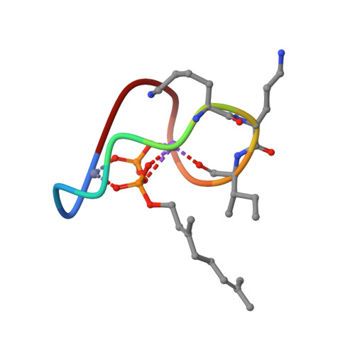
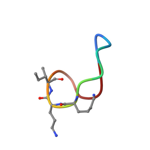
High-resolution crystal structure reveals molecular details of target recognition by bacitracin.

Economou, N.J.Cocklin, S.Loll, P.J.

(2013) Proc Natl Acad Sci U S A 110: 14207-14212

  • DOI: https://doi.org/10.1073/pnas.1308268110
  • Primary Citation of Related Structures:  
    4K7T

  • PubMed Abstract: 

    Bacitracin is a metalloantibiotic agent that is widely used as a medicine and feed additive. It interferes with bacterial cell-wall biosynthesis by binding undecaprenyl-pyrophosphate, a lipid carrier that serves as a critical intermediate in cell wall production. Despite bacitracin's broad use, the molecular details of its target recognition have not been elucidated. Here we report a crystal structure for the ternary complex of bacitracin A, zinc, and a geranyl-pyrophosphate ligand at a resolution of 1.1 Å. The antibiotic forms a compact structure that completely envelopes the ligand's pyrophosphate group, together with flanking zinc and sodium ions. The complex adopts a highly amphipathic conformation that offers clues to antibiotic function in the context of bacterial membranes. Bacitracin's efficient sequestration of its target represents a previously unseen mode for the recognition of lipid pyrophosphates, and suggests new directions for the design of next-generation antimicrobial agents.


  • Organizational Affiliation

    Department of Biochemistry and Molecular Biology, Drexel University College of Medicine, Philadelphia, PA 19102, USA.


Macromolecules

Find similar proteins by:  Sequence   |   3D Structure  

Entity ID: 1
MoleculeChains Sequence LengthOrganismDetailsImage
bacitracin A212Bacillus subtilisMutation(s): 0 
Entity Groups  
Sequence Clusters30% Identity50% Identity70% Identity90% Identity95% Identity100% Identity
Sequence Annotations
Expand
  • Reference Sequence
Biologically Interesting Molecules (External Reference) 1 Unique
Experimental Data & Validation

Experimental Data

  • Method: X-RAY DIFFRACTION
  • Resolution: 1.10 Å
  • R-Value Free:  0.149 (Depositor), 0.155 (DCC) 
  • R-Value Work:  0.127 (Depositor), 0.136 (DCC) 
  • R-Value Observed: 0.128 (Depositor) 
Space Group: P 62
Unit Cell:
Length ( Å )Angle ( ˚ )
a = 35.98α = 90
b = 35.98β = 90
c = 16.12γ = 120
Software Package:
Software NamePurpose
XSCALEdata scaling
PHENIXrefinement
PDB_EXTRACTdata extraction
ADSCdata collection
XDSdata reduction
PHENIXphasing

Structure Validation

View Full Validation Report


Ligand Structure Quality Assessment 

Created with Raphaël 2.3.0Worse 01 BetterLigand structure goodness of fit to experimental dataBest fitted GPPClick on this verticalbar to view details

Entry History 

Deposition Data

Revision History  (Full details and data files)

  • Version 1.0: 2013-08-14
    Type: Initial release
  • Version 1.1: 2013-09-18
    Changes: Database references
  • Version 1.2: 2013-10-16
    Changes: Derived calculations
  • Version 1.3: 2024-10-16
    Changes: Data collection, Database references, Derived calculations, Structure summary