4KVC

2H2 Fab fragment of immature Dengue virus


Experimental Data Snapshot

  • Method: X-RAY DIFFRACTION
  • Resolution: 2.31 Å
  • R-Value Free: 0.234 
  • R-Value Work: 0.179 
  • R-Value Observed: 0.182 

wwPDB Validation   3D Report Full Report


This is version 1.2 of the entry. See complete history


Literature

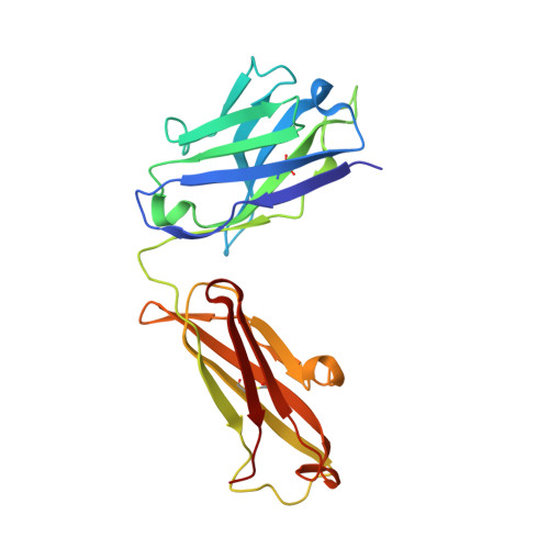
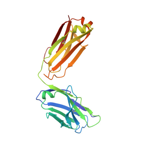
Obstruction of Dengue Virus Maturation by Fab Fragments of the 2H2 Antibody.

Wang, Z.Li, L.Pennington, J.G.Sheng, J.Yap, M.L.Plevka, P.Meng, G.Sun, L.Jiang, W.Rossmann, M.G.

(2013) J Virol 87: 8909-8915

  • DOI: https://doi.org/10.1128/JVI.00472-13
  • Primary Citation of Related Structures:  
    3J42, 4KVC

  • PubMed Abstract: 

    The 2H2 monoclonal antibody recognizes the precursor peptide on immature dengue virus and might therefore be a useful tool for investigating the conformational change that occurs when the immature virus enters an acidic environment. During dengue virus maturation, spiky, immature, noninfectious virions change their structure to form smooth-surfaced particles in the slightly acidic environment of the trans-Golgi network, thereby allowing cellular furin to cleave the precursor-membrane proteins. The dengue virions become fully infectious when they release the cleaved precursor peptide upon reaching the neutral-pH environment of the extracellular space. Here we report on the cryo-electron microscopy structures of the immature virus complexed with the 2H2 antigen binding fragments (Fab) at different concentrations and under various pH conditions. At neutral pH and a high concentration of Fab molecules, three Fab molecules bind to three precursor-membrane proteins on each spike of the immature virus. However, at a low concentration of Fab molecules and pH 7.0, only two Fab molecules bind to each spike. Changing to a slightly acidic pH caused no detectable change of structure for the sample with a high Fab concentration but caused severe structural damage to the low-concentration sample. Therefore, the 2H2 Fab inhibits the maturation process of immature dengue virus when Fab molecules are present at a high concentration, because the three Fab molecules on each spike hold the precursor-membrane molecules together, thereby inhibiting the normal conformational change that occurs during maturation.


  • Organizational Affiliation

    Department of Biological Sciences, Purdue University, West Lafayette, Indiana, USA.


Macromolecules
Find similar proteins by:  (by identity cutoff)  |  3D Structure
Entity ID: 1
MoleculeChains Sequence LengthOrganismDetailsImage
Ig heavy chain V region MOPC 21, Igh proteinA [auth H]220Mus musculusMutation(s): 0 
Gene Names: Igh-1aIgh
UniProt
Find proteins for P01863 (Mus musculus)
Explore P01863 
Go to UniProtKB:  P01863
Entity Groups  
Sequence Clusters30% Identity50% Identity70% Identity90% Identity95% Identity100% Identity
UniProt GroupP01863
Sequence Annotations
Expand
  • Reference Sequence
Find similar proteins by:  (by identity cutoff)  |  3D Structure
Entity ID: 2
MoleculeChains Sequence LengthOrganismDetailsImage
Ig kappa chain V-V region MOPC 21, Anti-colorectal carcinoma light chainB [auth L]212Mus musculusMutation(s): 0 
Gene Names: Gm16939
UniProt
Find proteins for P01837 (Mus musculus)
Explore P01837 
Go to UniProtKB:  P01837
Find proteins for P01634 (Mus musculus)
Explore P01634 
Go to UniProtKB:  P01634
Entity Groups  
Sequence Clusters30% Identity50% Identity70% Identity90% Identity95% Identity100% Identity
UniProt GroupsP01634P01837
Sequence Annotations
Expand
  • Reference Sequence
Experimental Data & Validation

Experimental Data

  • Method: X-RAY DIFFRACTION
  • Resolution: 2.31 Å
  • R-Value Free: 0.234 
  • R-Value Work: 0.179 
  • R-Value Observed: 0.182 
  • Space Group: P 2 21 21
Unit Cell:
Length ( Å )Angle ( ˚ )
a = 51.554α = 90
b = 81.711β = 90
c = 85.306γ = 90
Software Package:
Software NamePurpose
JBluIce-EPICSdata collection
MOLREPphasing
PHENIXrefinement
HKL-2000data reduction
HKL-2000data scaling

Structure Validation

View Full Validation Report


Entry History 

Deposition Data

Revision History  (Full details and data files)

  • Version 1.0: 2013-07-24
    Type: Initial release
  • Version 1.1: 2013-08-21
    Changes: Database references
  • Version 1.2: 2017-08-02
    Changes: Refinement description, Source and taxonomy