4O1W

Crystal Structure of Colwellia psychrerythraea cytochrome c


Experimental Data Snapshot

  • Method: X-RAY DIFFRACTION
  • Resolution: 2.00 Å
  • R-Value Free: 0.217 
  • R-Value Work: 0.170 
  • R-Value Observed: 0.172 

wwPDB Validation   3D Report Full Report


Ligand Structure Quality Assessment 


This is version 2.1 of the entry. See complete history


Literature

The structure of ferricytochrome c552 from the psychrophilic marine bacterium Colwellia psychrerythraea 34H.

Harvilla, P.B.Wolcott, H.N.Magyar, J.S.

(2014) Metallomics 6: 1126-1130

  • DOI: https://doi.org/10.1039/c4mt00045e
  • Primary Citation of Related Structures:  
    4O1W

  • PubMed Abstract: 

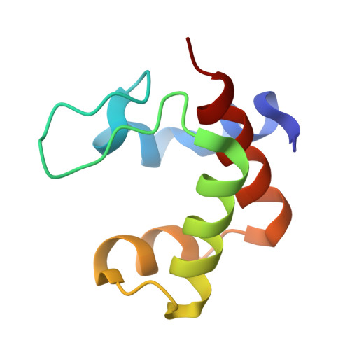
    Approximately 40% of all proteins are metalloproteins, and approximately 80% of Earth's ecosystems are at temperatures ≤5 °C, including 90% of the global ocean. Thus, an essential aspect of marine metallobiochemistry is an understanding of the structure, dynamics, and mechanisms of cold adaptation of metalloproteins from marine microorganisms. Here, the molecular structure of the electron-transfer protein cytochrome c552 from the psychrophilic marine bacterium Colwellia psychrerythraea 34H has been determined by X-ray crystallography (PDB: ). The structure is highly superimposable with that of the homologous cytochrome from the mesophile Marinobacter hydrocarbonoclasticus. Based on structural analysis and comparison of psychrophilic, psychrotolerant, and mesophilic sequences, a methionine-based ligand-substitution mechanism for psychrophilic protein stabilization is proposed.


  • Organizational Affiliation

    Department of Chemistry, Barnard College, Columbia University, 3009 Broadway, New York NY 10027, USA. jmagyar@barnard.edu.


Macromolecules
Find similar proteins by:  (by identity cutoff)  |  3D Structure
Entity ID: 1
MoleculeChains Sequence LengthOrganismDetailsImage
Cytochrome c552
A, B, C, D, E
A, B, C, D, E, F
79Colwellia psychrerythraea 34HMutation(s): 0 
Gene Names: CPS_0313
UniProt
Find proteins for Q48A34 (Colwellia psychrerythraea (strain 34H / ATCC BAA-681))
Explore Q48A34 
Go to UniProtKB:  Q48A34
Entity Groups  
Sequence Clusters30% Identity50% Identity70% Identity90% Identity95% Identity100% Identity
UniProt GroupQ48A34
Sequence Annotations
Expand
  • Reference Sequence
Small Molecules
Ligands 3 Unique
IDChains Name / Formula / InChI Key2D Diagram3D Interactions
HEC
Query on HEC

Download Ideal Coordinates CCD File 
G [auth A]
H [auth B]
J [auth C]
K [auth D]
M [auth E]
G [auth A],
H [auth B],
J [auth C],
K [auth D],
M [auth E],
O [auth F]
HEME C
C34 H34 Fe N4 O4
HXQIYSLZKNYNMH-LJNAALQVSA-N
PEG
Query on PEG

Download Ideal Coordinates CCD File 
I [auth B],
N [auth E]
DI(HYDROXYETHYL)ETHER
C4 H10 O3
MTHSVFCYNBDYFN-UHFFFAOYSA-N
SO4
Query on SO4

Download Ideal Coordinates CCD File 
L [auth D]SULFATE ION
O4 S
QAOWNCQODCNURD-UHFFFAOYSA-L
Experimental Data & Validation

Experimental Data

  • Method: X-RAY DIFFRACTION
  • Resolution: 2.00 Å
  • R-Value Free: 0.217 
  • R-Value Work: 0.170 
  • R-Value Observed: 0.172 
  • Space Group: C 1 2 1
Unit Cell:
Length ( Å )Angle ( ˚ )
a = 130.28α = 90
b = 45.043β = 90.85
c = 87.574γ = 90
Software Package:
Software NamePurpose
MAR345dtbdata collection
PHASERphasing
PHENIXrefinement
DENZOdata reduction
SCALEPACKdata scaling

Structure Validation

View Full Validation Report


Ligand Structure Quality Assessment 


Entry History 

Deposition Data

Revision History  (Full details and data files)

  • Version 1.0: 2014-04-23
    Type: Initial release
  • Version 1.1: 2014-05-28
    Changes: Database references
  • Version 1.2: 2014-06-18
    Changes: Database references
  • Version 2.0: 2021-03-10
    Changes: Atomic model, Data collection, Derived calculations, Non-polymer description, Structure summary
  • Version 2.1: 2023-09-20
    Changes: Data collection, Database references, Refinement description