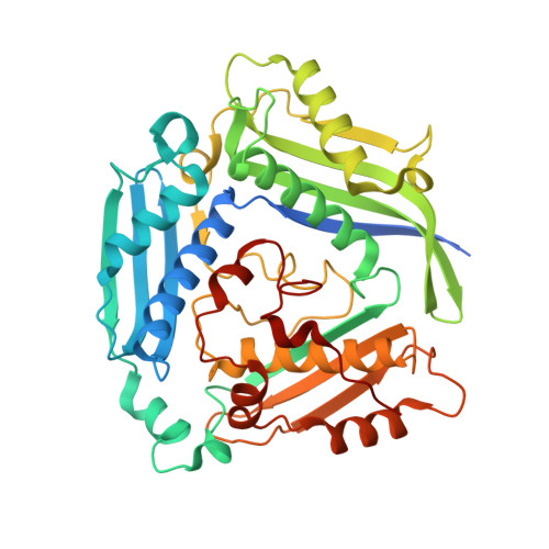
Crystal structure of a putative S-adenosylmethionine synthetase from Cryptosporidium hominis in complex with S-adenosyl-methionine
Seattle Structural Genomics Center for Infectious Disease (SSGCID), Abendroth, J., Arakaki, T., Lorimer, D., Edewars, T.E.To be published.