4OKM

Selinadiene Synthase apo and in complex with diphosphate


Experimental Data Snapshot

  • Method: X-RAY DIFFRACTION
  • Resolution: 2.10 Å
  • R-Value Free: 0.203 
  • R-Value Work: 0.157 
  • R-Value Observed: 0.159 

wwPDB Validation   3D Report Full Report


This is version 1.4 of the entry. See complete history


Literature

Induced-fit mechanism in class i terpene cyclases.

Baer, P.Rabe, P.Fischer, K.Citron, C.A.Klapschinski, T.A.Groll, M.Dickschat, J.S.

(2014) Angew Chem Int Ed Engl 53: 7652-7656

  • DOI: https://doi.org/10.1002/anie.201403648
  • Primary Citation of Related Structures:  
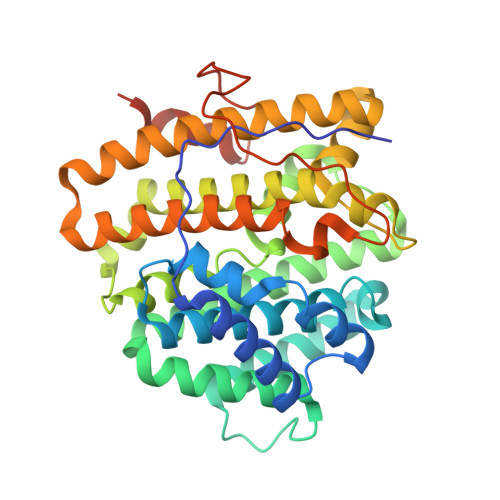
    4OKM, 4OKZ

  • PubMed Abstract: 

    We present crystallographic and functional data of selina-4(15),7(11)-diene synthase (SdS) from Streptomyces pristinaespiralis in its open and closed (ligand-bound) conformation. We could identify an induced-fit mechanism by elucidating a rearrangement of the G1/2 helix-break motif upon substrate binding. This rearrangement highlights a novel effector triad comprising the pyrophosphate sensor Arg178, the linker Asp181, and the effector Gly182-O. This structural motif is strictly conserved in class I terpene cyclases from bacteria, fungi, and plants, including epi-isozizaene synthase (3KB9), aristolochene synthase (4KUX), bornyl diphosphate synthase (1N20), limonene synthase (2ONG), 5-epi-aristolochene synthase (5EAT), and taxa-4(5),11(12)-diene synthase (3P5R). An elaborate structure-based mutagenesis in combination with analysis of the distinct product spectra confirmed the mechanistic models of carbocation formation and stabilization in SdS.


  • Organizational Affiliation

    Center for Integrated Protein Science at the Department Chemie, Lehrstuhl für Biochemie, TU München (Germany).


Macromolecules
Find similar proteins by:  (by identity cutoff)  |  3D Structure
Entity ID: 1
MoleculeChains Sequence LengthOrganismDetailsImage
Terpene synthase metal-binding domain-containing protein
A, B, C, D
365Streptomyces pristinaespiralis ATCC 25486Mutation(s): 0 
Gene Names: SSDG_02809ZP_06911744
EC: 2.5.1.21
UniProt
Find proteins for B5HDJ6 (Streptomyces pristinaespiralis (strain ATCC 25486 / DSM 40338 / CBS 914.69 / JCM 4507 / NBRC 13074 / NRRL 2958 / 5647))
Explore B5HDJ6 
Go to UniProtKB:  B5HDJ6
Entity Groups  
Sequence Clusters30% Identity50% Identity70% Identity90% Identity95% Identity100% Identity
UniProt GroupB5HDJ6
Sequence Annotations
Expand
  • Reference Sequence
Small Molecules
Ligands 2 Unique
IDChains Name / Formula / InChI Key2D Diagram3D Interactions
PPV
Query on PPV

Download Ideal Coordinates CCD File 
H [auth A],
L [auth B],
P [auth C]
PYROPHOSPHATE
H4 O7 P2
XPPKVPWEQAFLFU-UHFFFAOYSA-N
MG
Query on MG

Download Ideal Coordinates CCD File 
E [auth A]
F [auth A]
G [auth A]
I [auth B]
J [auth B]
E [auth A],
F [auth A],
G [auth A],
I [auth B],
J [auth B],
K [auth B],
M [auth C],
N [auth C],
O [auth C]
MAGNESIUM ION
Mg
JLVVSXFLKOJNIY-UHFFFAOYSA-N
Experimental Data & Validation

Experimental Data

  • Method: X-RAY DIFFRACTION
  • Resolution: 2.10 Å
  • R-Value Free: 0.203 
  • R-Value Work: 0.157 
  • R-Value Observed: 0.159 
  • Space Group: P 21 21 21
Unit Cell:
Length ( Å )Angle ( ˚ )
a = 74.78α = 90
b = 119.09β = 90
c = 186.1γ = 90
Software Package:
Software NamePurpose
XDSdata scaling
PHASERphasing
REFMACrefinement
XDSdata reduction
XSCALEdata scaling

Structure Validation

View Full Validation Report


Entry History 

Deposition Data

Revision History  (Full details and data files)

  • Version 1.0: 2014-06-11
    Type: Initial release
  • Version 1.1: 2014-07-02
    Changes: Database references
  • Version 1.2: 2014-08-06
    Changes: Database references
  • Version 1.3: 2024-02-28
    Changes: Data collection, Database references, Derived calculations
  • Version 1.4: 2024-04-03
    Changes: Refinement description