4R34

X-ray structure of the tryptophan lyase NosL with Tryptophan, 5'-deoxyadenosine and methionine bound


Experimental Data Snapshot

  • Method: X-RAY DIFFRACTION
  • Resolution: 1.80 Å
  • R-Value Free: 0.199 
  • R-Value Work: 0.172 
  • R-Value Observed: 0.174 

wwPDB Validation   3D Report Full Report


Ligand Structure Quality Assessment 


This is version 1.3 of the entry. See complete history


Literature

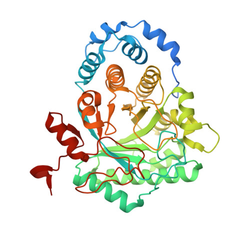
Crystal Structure of Tryptophan Lyase (NosL): Evidence for Radical Formation at the Amino Group of Tryptophan.

Nicolet, Y.Zeppieri, L.Amara, P.Fontecilla-Camps, J.C.

(2014) Angew Chem Int Ed Engl 53: 11840-11844

  • DOI: https://doi.org/10.1002/anie.201407320
  • Primary Citation of Related Structures:  
    4R33, 4R34

  • PubMed Abstract: 

    Streptomyces actuosus tryptophan lyase (NosL) is a radical SAM enzyme which catalyzes the synthesis of 3-methyl-2-indolic acid, a precursor in the synthesis of the promising antibiotic nosiheptide. The reaction involves cleavage of the tryptophan Cα-Cβ bond and recombination of the amino-acid-derived -COOH fragment at the indole ring. Reported herein is the 1.8 Å resolution crystal structure of NosL complexed with its substrate. Unexpectedly, only one of the tryptophan amino hydrogen atoms is optimally placed for H abstraction by the SAM-derived 5'-deoxyadenosyl radical. This orientation, in turn, rules out the previously proposed delocalized indole radical as the species which undergoes Cα-Cβ bond cleavage. Instead, stereochemical considerations indicate that the reactive intermediate is a (·)NH tryptophanyl radical. A structure-based amino acid sequence comparison of NosL with the tyrosine lyases ThiH and HydG strongly suggests that an equivalent (·)NH radical operates in the latter enzymes.


  • Organizational Affiliation

    Metalloproteins Unit, Institut de Biologie Structurale, UMR5075, CEA, CNRS, Université Grenoble-Alpes, 71, avenue des Martyrs, CS 10090, 38044 Grenoble cedex 9 (France). yvain.nicolet@ibs.fr.


Macromolecules
Find similar proteins by:  (by identity cutoff)  |  3D Structure
Entity ID: 1
MoleculeChains Sequence LengthOrganismDetailsImage
NosL
A, B
420Streptomyces actuosusMutation(s): 0 
Gene Names: nosL
UniProt
Find proteins for C6FX51 (Streptomyces actuosus)
Explore C6FX51 
Go to UniProtKB:  C6FX51
Entity Groups  
Sequence Clusters30% Identity50% Identity70% Identity90% Identity95% Identity100% Identity
UniProt GroupC6FX51
Sequence Annotations
Expand
  • Reference Sequence
Small Molecules
Ligands 9 Unique
IDChains Name / Formula / InChI Key2D Diagram3D Interactions
SAH
Query on SAH

Download Ideal Coordinates CCD File 
E [auth A],
O [auth B]
S-ADENOSYL-L-HOMOCYSTEINE
C14 H20 N6 O5 S
ZJUKTBDSGOFHSH-WFMPWKQPSA-N
SF4
Query on SF4

Download Ideal Coordinates CCD File 
F [auth A],
P [auth B]
IRON/SULFUR CLUSTER
Fe4 S4
LJBDFODJNLIPKO-UHFFFAOYSA-N
5AD
Query on 5AD

Download Ideal Coordinates CCD File 
D [auth A],
N [auth B]
5'-DEOXYADENOSINE
C10 H13 N5 O3
XGYIMTFOTBMPFP-KQYNXXCUSA-N
TRP
Query on TRP

Download Ideal Coordinates CCD File 
G [auth A],
Q [auth B]
TRYPTOPHAN
C11 H12 N2 O2
QIVBCDIJIAJPQS-VIFPVBQESA-N
MET
Query on MET

Download Ideal Coordinates CCD File 
C [auth A],
M [auth B]
METHIONINE
C5 H11 N O2 S
FFEARJCKVFRZRR-BYPYZUCNSA-N
GOL
Query on GOL

Download Ideal Coordinates CCD File 
H [auth A],
R [auth B],
S [auth B],
T [auth B]
GLYCEROL
C3 H8 O3
PEDCQBHIVMGVHV-UHFFFAOYSA-N
BR
Query on BR

Download Ideal Coordinates CCD File 
I [auth A]
J [auth A]
K [auth A]
U [auth B]
V [auth B]
I [auth A],
J [auth A],
K [auth A],
U [auth B],
V [auth B],
W [auth B],
Z [auth B]
BROMIDE ION
Br
CPELXLSAUQHCOX-UHFFFAOYSA-M
ZN
Query on ZN

Download Ideal Coordinates CCD File 
Y [auth B]ZINC ION
Zn
PTFCDOFLOPIGGS-UHFFFAOYSA-N
NA
Query on NA

Download Ideal Coordinates CCD File 
L [auth A],
X [auth B]
SODIUM ION
Na
FKNQFGJONOIPTF-UHFFFAOYSA-N
Experimental Data & Validation

Experimental Data

  • Method: X-RAY DIFFRACTION
  • Resolution: 1.80 Å
  • R-Value Free: 0.199 
  • R-Value Work: 0.172 
  • R-Value Observed: 0.174 
  • Space Group: P 1 2 1
Unit Cell:
Length ( Å )Angle ( ˚ )
a = 94.15α = 90
b = 47.07β = 108.99
c = 113.92γ = 90
Software Package:
Software NamePurpose
ADSCdata collection
PHENIXrefinement
XDSdata reduction
XSCALEdata scaling

Structure Validation

View Full Validation Report


Ligand Structure Quality Assessment 


Entry History 

Deposition Data

Revision History  (Full details and data files)

  • Version 1.0: 2014-09-17
    Type: Initial release
  • Version 1.1: 2014-11-12
    Changes: Database references
  • Version 1.2: 2017-11-22
    Changes: Refinement description
  • Version 1.3: 2024-02-28
    Changes: Data collection, Database references, Derived calculations