


Crystal Structure, Stability and siRNA Activity of Phosphorodithioate-Modified RNAs
Pallan, P.S., Yang, X., Sierant, M., Abeydeera, N.D., Hassell, T., Martinez, C., Janicka, M., Nawrot, B., Egli, M.To be published.
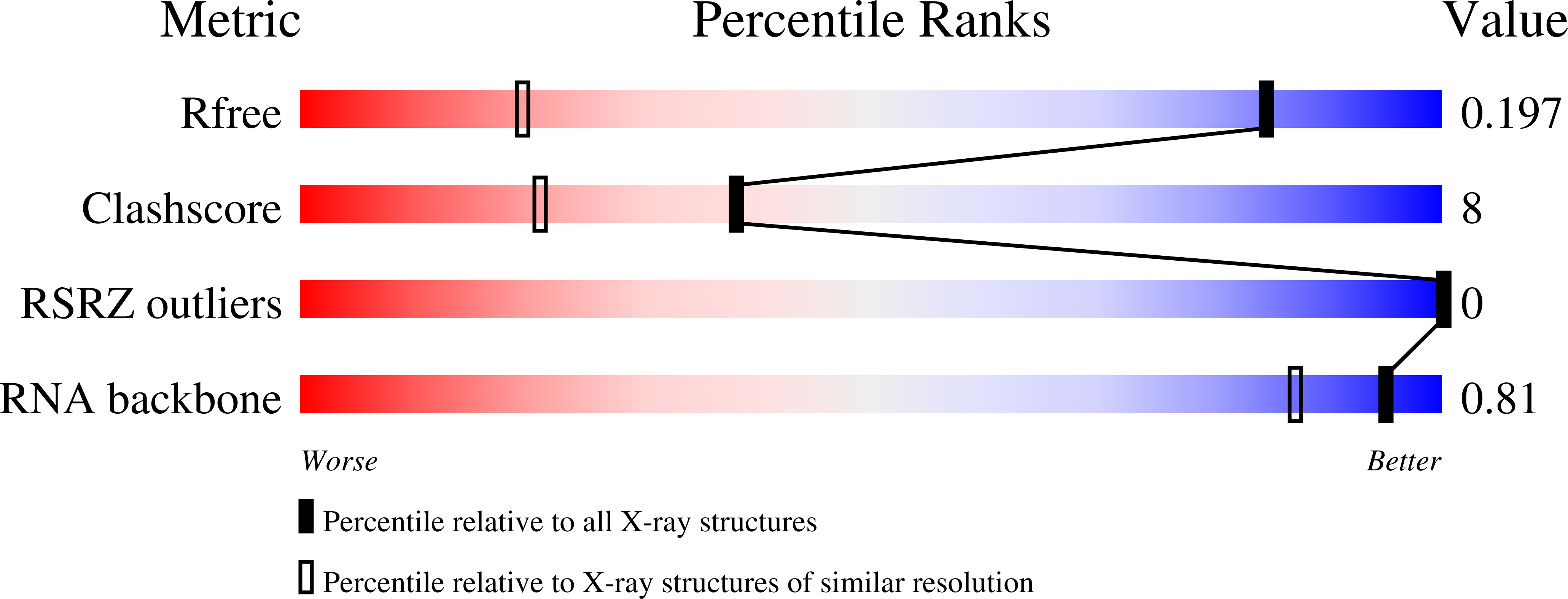
Experimental Data Snapshot
wwPDB Validation 3D Report Full Report
Find similar nucleic acids by: Sequence | 3D Structure
Entity ID: 1 | |||||
|---|---|---|---|---|---|
| Molecule | Chains | Length | Organism | Image | |
| 5'-R(*CP*GP*CP*(2SG)P*AP*AP*UP*UP*AP*GP*CP*G)-3' | 12 | synthetic construct | 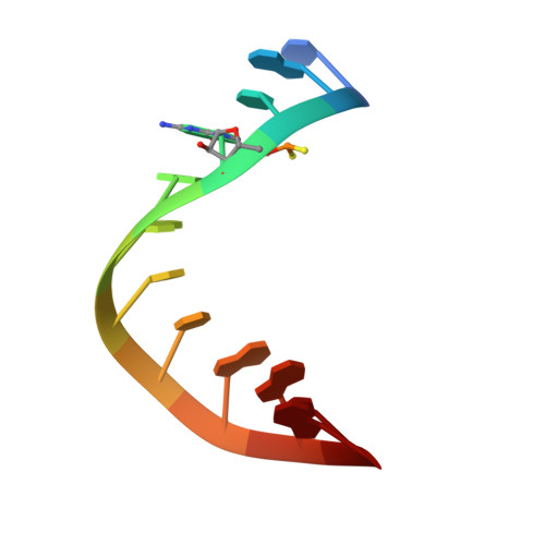 | ||
Sequence AnnotationsExpand | |||||
| |||||
| Ligands 1 Unique | |||||
|---|---|---|---|---|---|
| ID | Chains | Name / Formula / InChI Key | 2D Diagram | 3D Interactions | |
| SR Query on SR | B [auth A], C [auth A] | STRONTIUM ION Sr PWYYWQHXAPXYMF-UHFFFAOYSA-N |  | ||
| Length ( Å ) | Angle ( ˚ ) |
|---|---|
| a = 40.932 | α = 90 |
| b = 35.015 | β = 128.8 |
| c = 31.873 | γ = 90 |
| Software Name | Purpose |
|---|---|
| MD2 | data collection |
| MOLREP | phasing |
| SHELXL-97 | refinement |
| HKL-2000 | data reduction |
| HKL-2000 | data scaling |
RCSB PDB (citation) is hosted by
RCSB PDB is a member of the

