4TM8

Crystal structure of 6-phosphogluconolactonase from Mycobacterium smegmatis N131D mutant


Experimental Data Snapshot

  • Method: X-RAY DIFFRACTION
  • Resolution: 1.81 Å
  • R-Value Free: 0.201 
  • R-Value Work: 0.175 
  • R-Value Observed: 0.176 

wwPDB Validation   3D Report Full Report


This is version 1.2 of the entry. See complete history


Literature

Enzyme repurposing of a hydrolase as an emergent peroxidase upon metal binding.

Fujieda, N.Schatti, J.Stuttfeld, E.Ohkubo, K.Maier, T.Fukuzumi, S.Ward, T.R.

(2015) Chem Sci 6: 4060-4065

  • DOI: https://doi.org/10.1039/c5sc01065a
  • Primary Citation of Related Structures:  
    4TM7, 4TM8

  • PubMed Abstract: 

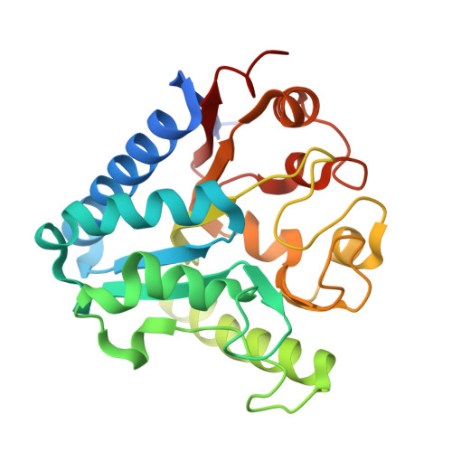
    As an alternative to Darwinian evolution relying on catalytic promiscuity, a protein may acquire auxiliary function upon metal binding, thus providing it with a novel catalytic machinery. Here we show that addition of cupric ions to a 6-phosphogluconolactonase 6-PGLac bearing a putative metal binding site leads to the emergence of peroxidase activity ( k cat 7.8 × 10 -2 s -1 , K M 1.1 × 10 -5 M). Both X-ray crystallographic and EPR data of the copper-loaded enzyme Cu·6-PGLac reveal a bis-histidine coordination site, located within a shallow binding pocket capable of accommodating the o -dianisidine substrate.


  • Organizational Affiliation

    Department of Chemistry , University of Basel , Spitalstrasse 51 , CH-4056 Basel , Switzerland . Email: fujieda@mls.eng.osaka-u.ac.jp ; Email: thomas.ward@unibas.ch.


Macromolecules
Find similar proteins by:  (by identity cutoff)  |  3D Structure
Entity ID: 1
MoleculeChains Sequence LengthOrganismDetailsImage
6-phosphogluconolactonase256Mycolicibacterium smegmatis MC2 155Mutation(s): 1 
Gene Names: pglMSMEG_3099MSMEI_3021
EC: 3.1.1.31
UniProt
Find proteins for A0QWX6 (Mycolicibacterium smegmatis (strain ATCC 700084 / mc(2)155))
Explore A0QWX6 
Go to UniProtKB:  A0QWX6
Entity Groups  
Sequence Clusters30% Identity50% Identity70% Identity90% Identity95% Identity100% Identity
UniProt GroupA0QWX6
Sequence Annotations
Expand
  • Reference Sequence
Small Molecules
Ligands 1 Unique
IDChains Name / Formula / InChI Key2D Diagram3D Interactions
SO4
Query on SO4

Download Ideal Coordinates CCD File 
B [auth A]SULFATE ION
O4 S
QAOWNCQODCNURD-UHFFFAOYSA-L
Experimental Data & Validation

Experimental Data

  • Method: X-RAY DIFFRACTION
  • Resolution: 1.81 Å
  • R-Value Free: 0.201 
  • R-Value Work: 0.175 
  • R-Value Observed: 0.176 
  • Space Group: I 41 2 2
Unit Cell:
Length ( Å )Angle ( ˚ )
a = 176.31α = 90
b = 176.31β = 90
c = 38.29γ = 90
Software Package:
Software NamePurpose
XSCALEdata scaling
PDB_EXTRACTdata extraction
PHENIXrefinement

Structure Validation

View Full Validation Report


Entry History 

Deposition Data

Revision History  (Full details and data files)

  • Version 1.0: 2015-06-03
    Type: Initial release
  • Version 1.1: 2018-01-24
    Changes: Database references, Derived calculations, Experimental preparation, Other, Source and taxonomy
  • Version 1.2: 2024-03-20
    Changes: Data collection, Database references, Refinement description