4XDC

Active semisynthetic [FeFe]-hydrogenase CpI with aza-dithiolato-bridged [2Fe] cofactor


Experimental Data Snapshot

  • Method: X-RAY DIFFRACTION
  • Resolution: 1.63 Å
  • R-Value Free: 0.176 
  • R-Value Work: 0.150 
  • R-Value Observed: 0.151 

wwPDB Validation   3D Report Full Report


Ligand Structure Quality Assessment 


This is version 1.3 of the entry. See complete history


Literature

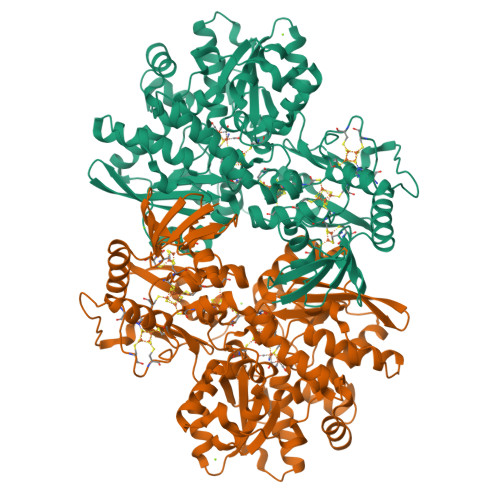
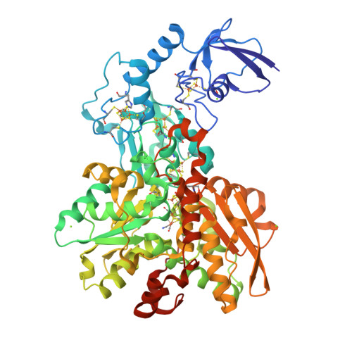
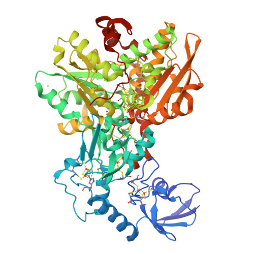
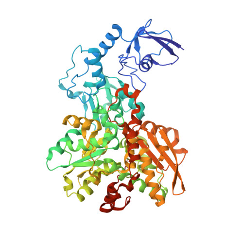
A structural view of synthetic cofactor integration into [FeFe]-hydrogenases.

Esselborn, J.Muraki, N.Klein, K.Engelbrecht, V.Metzler-Nolte, N.Apfel, U.P.Hofmann, E.Kurisu, G.Happe, T.

(2016) Chem Sci 7: 959-968

  • DOI: https://doi.org/10.1039/c5sc03397g
  • Primary Citation of Related Structures:  
    4XDC, 4XDD, 5BYQ, 5BYR, 5BYS

  • PubMed Abstract: 

    [FeFe]-hydrogenases are nature's fastest catalysts for the evolution or oxidation of hydrogen. Numerous synthetic model complexes for the [2Fe] subcluster (2Fe H ) of their active site are known, but so far none of these could compete with the enzymes. The complex Fe 2 [μ-(SCH 2 ) 2 X](CN) 2 (CO) 4 2- with X = NH was shown to integrate into the apo-form of [FeFe]-hydrogenases to yield a fully active enzyme. Here we report the first crystal structures of the apo-form of the bacterial [FeFe]-hydrogenase CpI from Clostridium pasteurianum at 1.60 Å and the active semisynthetic enzyme, CpI ADT , at 1.63 Å. The structures illustrate the significant changes in ligand coordination upon integration and activation of the [2Fe] complex. These changes are induced by a rigid 2Fe H cavity as revealed by the structure of apoCpI, which is remarkably similar to CpI ADT . Additionally we present the high resolution crystal structures of the semisynthetic bacterial [FeFe]-hydrogenases CpI PDT (X = CH 2 ), CpI ODT (X = O) and CpI SDT (X = S) with changes in the headgroup of the dithiolate bridge in the 2Fe H cofactor. The structures of these inactive enzymes demonstrate that the 2Fe H -subcluster and its protein environment remain largely unchanged when compared to the active enzyme CpI ADT . As the active site shows an open coordination site in all structures, the absence of catalytic activity is probably not caused by steric obstruction. This demonstrates that the chemical properties of the dithiolate bridge are essential for enzyme activity.


  • Organizational Affiliation

    AG Photobiotechnologie , Fakultät für Biologie und Biotechnologie , Ruhr-Universität Bochum , Universitätsstraße 150 , 44801 Bochum , Germany . Email: thomas.happe@rub.de.


Macromolecules
Find similar proteins by:  (by identity cutoff)  |  3D Structure
Entity ID: 1
MoleculeChains Sequence LengthOrganismDetailsImage
Iron hydrogenase 1
A, B
584Clostridium pasteurianumMutation(s): 0 
EC: 1.12.7.2
UniProt
Find proteins for P29166 (Clostridium pasteurianum)
Explore P29166 
Go to UniProtKB:  P29166
Entity Groups  
Sequence Clusters30% Identity50% Identity70% Identity90% Identity95% Identity100% Identity
UniProt GroupP29166
Sequence Annotations
Expand
  • Reference Sequence
Small Molecules
Ligands 4 Unique
IDChains Name / Formula / InChI Key2D Diagram3D Interactions
402
Query on 402

Download Ideal Coordinates CCD File 
C [auth A],
L [auth B]
dicarbonyl[bis(cyanide-kappaC)]-mu-(iminodimethanethiolatato-1kappaS:2kappaS)-mu-(oxomethylidene)diiron(2+)
C7 H5 Fe2 N3 O3 S2
LJPDYWPSPOWMIB-UHFFFAOYSA-N
SF4
Query on SF4

Download Ideal Coordinates CCD File 
D [auth A]
E [auth A]
F [auth A]
G [auth A]
M [auth B]
D [auth A],
E [auth A],
F [auth A],
G [auth A],
M [auth B],
N [auth B],
O [auth B],
P [auth B]
IRON/SULFUR CLUSTER
Fe4 S4
LJBDFODJNLIPKO-UHFFFAOYSA-N
FES
Query on FES

Download Ideal Coordinates CCD File 
H [auth A],
Q [auth B]
FE2/S2 (INORGANIC) CLUSTER
Fe2 S2
NIXDOXVAJZFRNF-UHFFFAOYSA-N
MG
Query on MG

Download Ideal Coordinates CCD File 
I [auth A],
J [auth A],
K [auth A],
R [auth B],
S [auth B]
MAGNESIUM ION
Mg
JLVVSXFLKOJNIY-UHFFFAOYSA-N
Experimental Data & Validation

Experimental Data

  • Method: X-RAY DIFFRACTION
  • Resolution: 1.63 Å
  • R-Value Free: 0.176 
  • R-Value Work: 0.150 
  • R-Value Observed: 0.151 
  • Space Group: P 1 21 1
Unit Cell:
Length ( Å )Angle ( ˚ )
a = 91.34α = 90
b = 73.65β = 97.28
c = 103.88γ = 90
Software Package:
Software NamePurpose
PHENIXrefinement
XDSdata reduction
PHASERphasing
XSCALEdata scaling

Structure Validation

View Full Validation Report


Ligand Structure Quality Assessment 


Entry History 

Deposition Data

Revision History  (Full details and data files)

  • Version 1.0: 2015-11-11
    Type: Initial release
  • Version 1.1: 2018-07-11
    Changes: Data collection, Database references
  • Version 1.2: 2019-10-02
    Changes: Data collection, Derived calculations
  • Version 1.3: 2024-01-10
    Changes: Data collection, Database references, Derived calculations, Refinement description