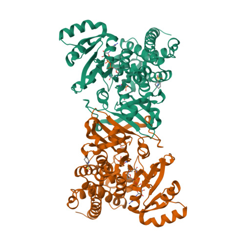
Crystal structures of the Moraxella catarrhalis DOX-P Reductoisomerase
Birkinshaw, R.W., Brady, R.L.To be published.
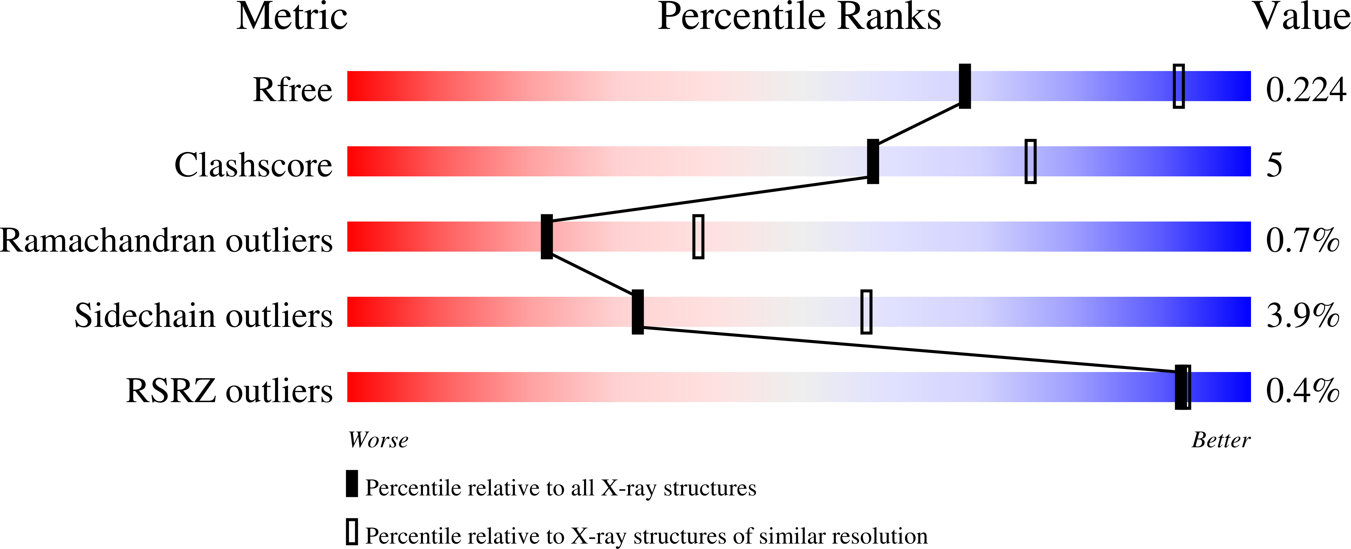
Experimental Data Snapshot
Entity ID: 1 | |||||
|---|---|---|---|---|---|
| Molecule | Chains | Sequence Length | Organism | Details | Image |
| 1-deoxy-D-xylulose 5-phosphate reductoisomerase | 415 | Moraxella catarrhalis | Mutation(s): 0 Gene Names: dxr, DR90_1378 EC: 1.1.1.267 | 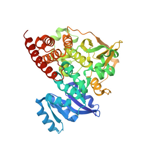 | |
Entity Groups | |||||
| Sequence Clusters | 30% Identity50% Identity70% Identity90% Identity95% Identity100% Identity | ||||
Sequence AnnotationsExpand | |||||
| |||||
| Ligands 5 Unique | |||||
|---|---|---|---|---|---|
| ID | Chains | Name / Formula / InChI Key | 2D Diagram | 3D Interactions | |
| NAD Query on NAD | C [auth A], H [auth B] | NICOTINAMIDE-ADENINE-DINUCLEOTIDE C21 H27 N7 O14 P2 BAWFJGJZGIEFAR-NNYOXOHSSA-N |  | ||
| NHE Query on NHE | G [auth A], L [auth B] | 2-[N-CYCLOHEXYLAMINO]ETHANE SULFONIC ACID C8 H17 N O3 S MKWKNSIESPFAQN-UHFFFAOYSA-N |  | ||
| FOM Query on FOM | E [auth A], J [auth B] | 3-[FORMYL(HYDROXY)AMINO]PROPYLPHOSPHONIC ACID C4 H10 N O5 P GJXWDTUCERCKIX-UHFFFAOYSA-N |  | ||
| GOL Query on GOL | F [auth A], K [auth B] | GLYCEROL C3 H8 O3 PEDCQBHIVMGVHV-UHFFFAOYSA-N |  | ||
| MG Query on MG | D [auth A], I [auth B] | MAGNESIUM ION Mg JLVVSXFLKOJNIY-UHFFFAOYSA-N |  | ||
| Length ( Å ) | Angle ( ˚ ) |
|---|---|
| a = 65.719 | α = 90 |
| b = 65.719 | β = 90 |
| c = 390.106 | γ = 120 |
| Software Name | Purpose |
|---|---|
| REFMAC | refinement |
| XDS | data reduction |
| SCALA | data scaling |
| PHASER | phasing |