5ACI

X-ray Structure of LPMO


Experimental Data Snapshot

  • Method: X-RAY DIFFRACTION
  • Resolution: 1.75 Å
  • R-Value Free: 0.195 
  • R-Value Work: 0.165 
  • R-Value Observed: 0.166 

wwPDB Validation   3D Report Full Report


Ligand Structure Quality Assessment 


This is version 2.1 of the entry. See complete history


Literature

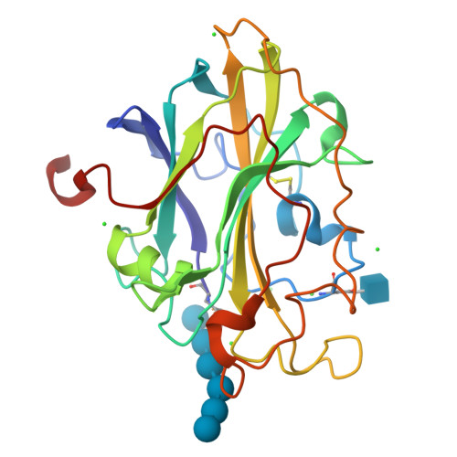
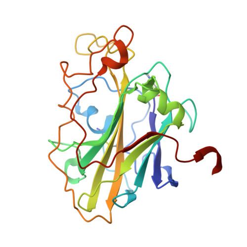
The molecular basis of polysaccharide cleavage by lytic polysaccharide monooxygenases.

Frandsen, K.E.Simmons, T.J.Dupree, P.Poulsen, J.C.Hemsworth, G.R.Ciano, L.Johnston, E.M.Tovborg, M.Johansen, K.S.von Freiesleben, P.Marmuse, L.Fort, S.Cottaz, S.Driguez, H.Henrissat, B.Lenfant, N.Tuna, F.Baldansuren, A.Davies, G.J.Lo Leggio, L.Walton, P.H.

(2016) Nat Chem Biol 12: 298-303

  • DOI: https://doi.org/10.1038/nchembio.2029
  • Primary Citation of Related Structures:  
    5ACF, 5ACG, 5ACH, 5ACI, 5ACJ

  • PubMed Abstract: 

    Lytic polysaccharide monooxygenases (LPMOs) are copper-containing enzymes that oxidatively break down recalcitrant polysaccharides such as cellulose and chitin. Since their discovery, LPMOs have become integral factors in the industrial utilization of biomass, especially in the sustainable generation of cellulosic bioethanol. We report here a structural determination of an LPMO-oligosaccharide complex, yielding detailed insights into the mechanism of action of these enzymes. Using a combination of structure and electron paramagnetic resonance spectroscopy, we reveal the means by which LPMOs interact with saccharide substrates. We further uncover electronic and structural features of the enzyme active site, showing how LPMOs orchestrate the reaction of oxygen with polysaccharide chains.


  • Organizational Affiliation

    Department of Chemistry, University of Copenhagen, Copenhagen, Denmark.


Macromolecules
Find similar proteins by:  (by identity cutoff)  |  3D Structure
Entity ID: 1
MoleculeChains Sequence LengthOrganismDetailsImage
LYTIC POLYSACCHARIDE MONOOXYGENASE235Panus similisMutation(s): 0 
UniProt
Find proteins for A0A0S2GKZ1 (Panus similis)
Explore A0A0S2GKZ1 
Go to UniProtKB:  A0A0S2GKZ1
Entity Groups  
Sequence Clusters30% Identity50% Identity70% Identity90% Identity95% Identity100% Identity
UniProt GroupA0A0S2GKZ1
Sequence Annotations
Expand
  • Reference Sequence
Oligosaccharides

Help

Entity ID: 2
MoleculeChains Length2D Diagram Glycosylation3D Interactions
beta-D-glucopyranose-(1-4)-beta-D-glucopyranose-(1-4)-beta-D-glucopyranose-(1-4)-beta-D-glucopyranose-(1-4)-beta-D-glucopyranose-(1-4)-beta-D-glucopyranose
B
6N/A
Glycosylation Resources
GlyTouCan:  G09454VW
GlyCosmos:  G09454VW
GlyGen:  G09454VW
Small Molecules
Ligands 3 Unique
IDChains Name / Formula / InChI Key2D Diagram3D Interactions
NAG
Query on NAG

Download Ideal Coordinates CCD File 
D [auth A]2-acetamido-2-deoxy-beta-D-glucopyranose
C8 H15 N O6
OVRNDRQMDRJTHS-FMDGEEDCSA-N
CU
Query on CU

Download Ideal Coordinates CCD File 
C [auth A]COPPER (II) ION
Cu
JPVYNHNXODAKFH-UHFFFAOYSA-N
CL
Query on CL

Download Ideal Coordinates CCD File 
E [auth A]
F [auth A]
G [auth A]
H [auth A]
I [auth A]
E [auth A],
F [auth A],
G [auth A],
H [auth A],
I [auth A],
J [auth A],
K [auth A]
CHLORIDE ION
Cl
VEXZGXHMUGYJMC-UHFFFAOYSA-M
Modified Residues  1 Unique
IDChains TypeFormula2D DiagramParent
HIC
Query on HIC
A
L-PEPTIDE LINKINGC7 H11 N3 O2HIS
Biologically Interesting Molecules (External Reference) 1 Unique
Experimental Data & Validation

Experimental Data

  • Method: X-RAY DIFFRACTION
  • Resolution: 1.75 Å
  • R-Value Free: 0.195 
  • R-Value Work: 0.165 
  • R-Value Observed: 0.166 
  • Space Group: P 41 3 2
Unit Cell:
Length ( Å )Angle ( ˚ )
a = 125.46α = 90
b = 125.46β = 90
c = 125.46γ = 90
Software Package:
Software NamePurpose
REFMACrefinement
XDSdata reduction
XSCALEdata scaling
MOLREPphasing

Structure Validation

View Full Validation Report


Ligand Structure Quality Assessment 


Entry History 

Deposition Data

Revision History  (Full details and data files)

  • Version 1.0: 2016-03-02
    Type: Initial release
  • Version 1.1: 2016-03-09
    Changes: Database references
  • Version 1.2: 2016-03-30
    Changes: Database references
  • Version 1.3: 2018-01-17
    Changes: Data collection, Database references, Structure summary
  • Version 2.0: 2020-07-29
    Type: Remediation
    Reason: Carbohydrate remediation
    Changes: Atomic model, Data collection, Derived calculations, Other, Structure summary
  • Version 2.1: 2024-01-10
    Changes: Data collection, Database references, Refinement description, Structure summary