5C04

Crystal structure of the F37H mutant AhpE from Mycobacterium tuberculosis


Experimental Data Snapshot

  • Method: X-RAY DIFFRACTION
  • Resolution: 1.45 Å
  • R-Value Free: 0.198 
  • R-Value Work: 0.166 
  • R-Value Observed: 0.167 

wwPDB Validation   3D Report Full Report


This is version 1.3 of the entry. See complete history


Literature

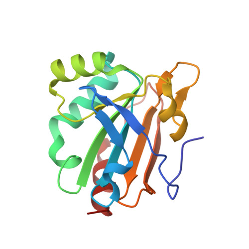
The active site architecture in peroxiredoxins: a case study on Mycobacterium tuberculosis AhpE.

Pedre, B.van Bergen, L.A.Pallo, A.Rosado, L.A.Dufe, V.T.Molle, I.V.Wahni, K.Erdogan, H.Alonso, M.Proft, F.D.Messens, J.

(2016) Chem Commun (Camb) 52: 10293-10296

  • DOI: https://doi.org/10.1039/c6cc02645a
  • Primary Citation of Related Structures:  
    4XIH, 5C04

  • PubMed Abstract: 

    Peroxiredoxins catalyze the reduction of peroxides, a process of vital importance to survive oxidative stress. A nucleophilic cysteine, also known as the peroxidatic cysteine, is responsible for this catalytic process. We used the Mycobacterium tuberculosis alkyl hydroperoxide reductase E (MtAhpE) as a model to investigate the effect of the chemical environment on the specificity of the reaction. Using an integrative structural (R116A - PDB ; F37H - PDB ), kinetic and computational approach, we explain the mutational effects of key residues in its environment. This study shows that the active site residues are specifically oriented to create an environment which selectively favours a reaction with peroxides.


  • Organizational Affiliation

    Structural Biology Research Center, Oxidative Stress Signaling lab, VIB, Pleinlaan 2, 1050 Brussels, Belgium. joris.messens@vib-vub.be and Brussels Center for Redox Biology, 1050 Brussels, Belgium and Structural Biology Brussels, Vrije Universiteit Brussel, 1050 Brussels, Belgium.


Macromolecules
Find similar proteins by:  (by identity cutoff)  |  3D Structure
Entity ID: 1
MoleculeChains Sequence LengthOrganismDetailsImage
Putative peroxiredoxin MT2298
A, B
153Mycobacterium tuberculosisMutation(s): 1 
Gene Names: MT2298
EC: 1.11.1.15
UniProt
Find proteins for P9WIE3 (Mycobacterium tuberculosis (strain ATCC 25618 / H37Rv))
Explore P9WIE3 
Go to UniProtKB:  P9WIE3
Entity Groups  
Sequence Clusters30% Identity50% Identity70% Identity90% Identity95% Identity100% Identity
UniProt GroupP9WIE3
Sequence Annotations
Expand
  • Reference Sequence
Experimental Data & Validation

Experimental Data

  • Method: X-RAY DIFFRACTION
  • Resolution: 1.45 Å
  • R-Value Free: 0.198 
  • R-Value Work: 0.166 
  • R-Value Observed: 0.167 
  • Space Group: P 43 21 2
Unit Cell:
Length ( Å )Angle ( ˚ )
a = 63.3α = 90
b = 63.3β = 90
c = 159.45γ = 90
Software Package:
Software NamePurpose
XDSdata reduction
XSCALEdata scaling
MOLREPphasing
Cootmodel building
REFMACrefinement
PDB_EXTRACTdata extraction

Structure Validation

View Full Validation Report


Entry History & Funding Information

Deposition Data


Funding OrganizationLocationGrant Number
Research Foundation - Flanders (FWO)BelgiumG.0305.12

Revision History  (Full details and data files)

  • Version 1.0: 2016-07-27
    Type: Initial release
  • Version 1.1: 2016-08-10
    Changes: Database references
  • Version 1.2: 2016-08-24
    Changes: Database references
  • Version 1.3: 2024-01-10
    Changes: Author supporting evidence, Data collection, Database references, Refinement description