5DM0 | pdb_00005dm0

Crystal structure of the plantazolicin methyltransferase BamL in complex with triazolic desmethylPZN analog and SAH


Experimental Data Snapshot

  • Method: X-RAY DIFFRACTION
  • Resolution: 1.75 Å
  • R-Value Free: 
    0.212 (Depositor), 0.210 (DCC) 
  • R-Value Work: 
    0.186 (Depositor), 0.190 (DCC) 
  • R-Value Observed: 
    0.187 (Depositor) 

wwPDB Validation   3D Report Full Report


Ligand Structure Quality Assessment 

Created with Raphaël 2.3.0Worse 01 BetterLigand structure goodness of fit to experimental dataBest fitted 5D8Click on this verticalbar to view detailsBest fitted SAHClick on this verticalbar to view details

This is version 1.2 of the entry. See complete history


Literature

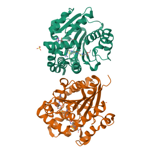
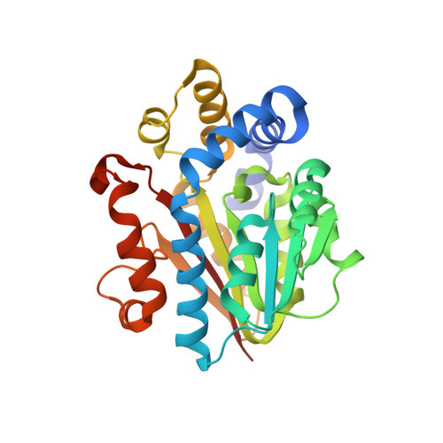
Insights into methyltransferase specificity and bioactivity of derivatives of the antibiotic plantazolicin.

Hao, Y.Blair, P.M.Sharma, A.Mitchell, D.A.Nair, S.K.

(2015) ACS Chem Biol 10: 1209-1216

  • DOI: https://doi.org/10.1021/cb501042a
  • Primary Citation of Related Structures:  
    5DLY, 5DM0, 5DM1, 5DM2, 5DM4

  • PubMed Abstract: 

    Peptide antibiotics represent a class of conformationally constrained natural products of growing pharmaceutical interest. Plantazolicin (PZN) is a linear, polyheterocyclic natural product with highly selective and potent activity against the anthrax-causing bacterium, Bacillus anthracis. The bioactivity of PZN is contingent on dimethylation of its N-terminal Arg residue by an S-adenosylmethionine-dependent methyltransferase. Here, we explore the substrate tolerances of two homologous PZN methyltransferases by carrying out kinetic analyses of the enzymes against a synthetic panel of truncated PZN analogs containing the N-terminal Arg residue. X-ray cocrystal structures of the PZN methyltransferases with each of these heterocycle-containing substrates provide a rationale for understanding the strict substrate specificity of these enzymes. Kinetic studies of structure-guided, site-specific variants allowed for the assignment of residues governing catalysis and substrate scope. Microbiological testing further revealed that upon dimethylation of the N-terminal Arg, a pentaheterocyclized PZN analog retained potent anti-B. anthracis activity, nearly equal to that of full-length PZN. These studies may be useful in the biosynthetic engineering of natural product analogs with different bioactivity profiles, as demonstrated by our identification of a truncated plantazolicin derivative that is active against methicillin-resistant Staphylococcus aureus (MRSA).


  • Organizational Affiliation

    Department of Biochemistry, University of Illinois at Urbana-Champaign, Urbana, IL 61801.


Macromolecules
Find similar proteins by:  (by identity cutoff)  |  3D Structure
Entity ID: 1
MoleculeChains Sequence LengthOrganismDetailsImage
plantazolicin methyltransferase BamL
A, B
272Bacillus velezensis FZB42Mutation(s): 0 
Gene Names: RBAM_007500
UniProt
Find proteins for A7Z2A9 (Bacillus velezensis (strain DSM 23117 / BGSC 10A6 / LMG 26770 / FZB42))
Explore A7Z2A9 
Go to UniProtKB:  A7Z2A9
Entity Groups  
Sequence Clusters30% Identity50% Identity70% Identity90% Identity95% Identity100% Identity
UniProt GroupA7Z2A9
Sequence Annotations
Expand
  • Reference Sequence
Small Molecules
Ligands 3 Unique
IDChains Name / Formula / InChI Key2D Diagram3D Interactions
5D8
Query on 5D8

Download Ideal Coordinates CCD File 
E [auth A],
H [auth B]
ethyl 2-(2-{2-[(1S)-1-amino-4-carbamimidamidobutyl]-1,3-thiazol-4-yl}-5-methyl-1,3-oxazol-4-yl)-1,3-thiazole-4-carboxylate
C18 H23 N7 O3 S2
LDXVRBOROSJVJC-JTQLQIEISA-N
SAH
Query on SAH

Download Ideal Coordinates CCD File 
D [auth A],
G [auth B]
S-ADENOSYL-L-HOMOCYSTEINE
C14 H20 N6 O5 S
ZJUKTBDSGOFHSH-WFMPWKQPSA-N
SO4
Query on SO4

Download Ideal Coordinates CCD File 
C [auth A],
F [auth B]
SULFATE ION
O4 S
QAOWNCQODCNURD-UHFFFAOYSA-L
Experimental Data & Validation

Experimental Data

  • Method: X-RAY DIFFRACTION
  • Resolution: 1.75 Å
  • R-Value Free:  0.212 (Depositor), 0.210 (DCC) 
  • R-Value Work:  0.186 (Depositor), 0.190 (DCC) 
  • R-Value Observed: 0.187 (Depositor) 
Space Group: P 21 21 21
Unit Cell:
Length ( Å )Angle ( ˚ )
a = 48.839α = 90
b = 96.501β = 90
c = 132.646γ = 90
Software Package:
Software NamePurpose
REFMACrefinement
HKL-2000data scaling
PHENIXmodel building
Cootmodel building

Structure Validation

View Full Validation Report


Ligand Structure Quality Assessment 

Created with Raphaël 2.3.0Worse 01 BetterLigand structure goodness of fit to experimental dataBest fitted 5D8Click on this verticalbar to view detailsBest fitted SAHClick on this verticalbar to view details

Entry History 

Deposition Data

Revision History  (Full details and data files)

  • Version 1.0: 2015-09-23
    Type: Initial release
  • Version 1.1: 2017-11-22
    Changes: Derived calculations, Refinement description
  • Version 1.2: 2024-03-06
    Changes: Data collection, Database references