5H0U

Crystal structure of the catalytic domain of membrane type 1 matrix metalloproteinase


Experimental Data Snapshot

  • Method: X-RAY DIFFRACTION
  • Resolution: 2.24 Å
  • R-Value Free: 0.254 
  • R-Value Work: 0.195 
  • R-Value Observed: 0.198 

wwPDB Validation   3D Report Full Report


This is version 1.3 of the entry. See complete history


Literature

Solvent water interactions within the active site of the membrane type I matrix metalloproteinase.

Decaneto, E.Vasilevskaya, T.Kutin, Y.Ogata, H.Grossman, M.Sagi, I.Havenith, M.Lubitz, W.Thiel, W.Cox, N.

(2017) Phys Chem Chem Phys 19: 30316-30331

  • DOI: https://doi.org/10.1039/c7cp05572b
  • Primary Citation of Related Structures:  
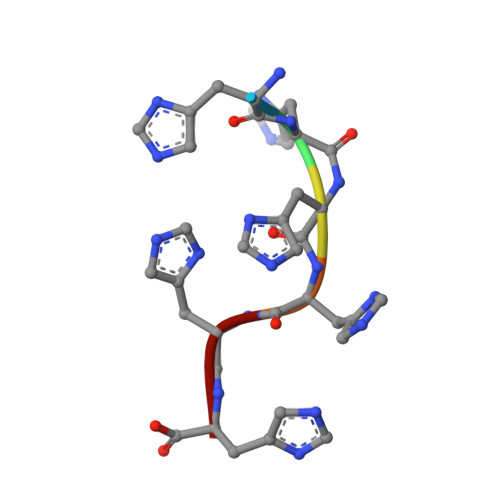
    5H0U

  • PubMed Abstract: 

    Matrix metalloproteinases (MMP) are an important family of proteases which catalyze the degradation of extracellular matrix components. While the mechanism of peptide cleavage is well established, the process of enzyme regeneration, which represents the rate limiting step of the catalytic cycle, remains unresolved. This step involves the loss of the newly formed N-terminus (amine) and C-terminus (carboxylate) protein fragments from the site of catalysis coupled with the inclusion of one or more solvent waters. Here we report a novel crystal structure of membrane type I MMP (MT1-MMP or MMP-14), which includes a small peptide bound at the catalytic Zn site via its C-terminus. This structure models the initial product state formed immediately after peptide cleavage but before the final proton transfer to the bound amine; the amine is not present in our system and as such proton transfer cannot occur. Modeling of the protein, including earlier structural data of Bertini and coworkers [I. Bertini, et al., Angew. Chem., Int. Ed., 2006, 45, 7952-7955], suggests that the C-terminus of the peptide is positioned to form an H-bond network to the amine site, which is mediated by a single oxygen of the functionally important Glu240 residue, facilitating efficient proton transfer. Additional quantum chemical calculations complemented with magneto-optical and magnetic resonance spectroscopies clarify the role of two additional, non-catalytic first coordination sphere waters identified in the crystal structure. One of these auxiliary waters acts to stabilize key intermediates of the reaction, while the second is proposed to facilitate C-fragment release, triggered by protonation of the amine. Together these results complete the enzymatic cycle of MMPs and provide new design criteria for inhibitors with improved efficacy.


  • Organizational Affiliation

    Max Planck Institute for Chemical Energy Conversion, Stiftstraße. 34-36, D-45470, Mülheim an der Ruhr, Germany. nicholas.cox@cec.mpg.de.


Macromolecules
Find similar proteins by:  (by identity cutoff)  |  3D Structure
Entity ID: 1
MoleculeChains Sequence LengthOrganismDetailsImage
Matrix metalloproteinase-14170Homo sapiensMutation(s): 0 
Gene Names: MMP14
EC: 3.4.24.80
UniProt & NIH Common Fund Data Resources
Find proteins for P50281 (Homo sapiens)
Explore P50281 
Go to UniProtKB:  P50281
PHAROS:  P50281
GTEx:  ENSG00000157227 
Entity Groups  
Sequence Clusters30% Identity50% Identity70% Identity90% Identity95% Identity100% Identity
UniProt GroupP50281
Sequence Annotations
Expand
  • Reference Sequence

Find similar proteins by:  Sequence   |   3D Structure  

Entity ID: 2
MoleculeChains Sequence LengthOrganismDetailsImage
HIS-HIS-HIS-HIS-HIS-HIS6unidentifiedMutation(s): 0 
Sequence Annotations
Expand
  • Reference Sequence
Small Molecules
Ligands 4 Unique
IDChains Name / Formula / InChI Key2D Diagram3D Interactions
EPE
Query on EPE

Download Ideal Coordinates CCD File 
G [auth A]4-(2-HYDROXYETHYL)-1-PIPERAZINE ETHANESULFONIC ACID
C8 H18 N2 O4 S
JKMHFZQWWAIEOD-UHFFFAOYSA-N
GOL
Query on GOL

Download Ideal Coordinates CCD File 
H [auth A]
I [auth A]
J [auth A]
K [auth A]
L [auth A]
H [auth A],
I [auth A],
J [auth A],
K [auth A],
L [auth A],
M [auth A],
N [auth A],
O [auth A]
GLYCEROL
C3 H8 O3
PEDCQBHIVMGVHV-UHFFFAOYSA-N
ZN
Query on ZN

Download Ideal Coordinates CCD File 
D [auth A],
E [auth A],
P [auth A]
ZINC ION
Zn
PTFCDOFLOPIGGS-UHFFFAOYSA-N
CA
Query on CA

Download Ideal Coordinates CCD File 
C [auth A],
F [auth A]
CALCIUM ION
Ca
BHPQYMZQTOCNFJ-UHFFFAOYSA-N
Experimental Data & Validation

Experimental Data

  • Method: X-RAY DIFFRACTION
  • Resolution: 2.24 Å
  • R-Value Free: 0.254 
  • R-Value Work: 0.195 
  • R-Value Observed: 0.198 
  • Space Group: P 43 21 2
Unit Cell:
Length ( Å )Angle ( ˚ )
a = 62.995α = 90
b = 62.995β = 90
c = 122.62γ = 90
Software Package:
Software NamePurpose
PHENIXrefinement
XDSdata reduction
XSCALEdata scaling
MOLREPphasing

Structure Validation

View Full Validation Report


Entry History 

Deposition Data

Revision History  (Full details and data files)

  • Version 1.0: 2017-10-04
    Type: Initial release
  • Version 1.1: 2017-10-11
    Changes: Database references
  • Version 1.2: 2017-11-29
    Changes: Database references
  • Version 1.3: 2024-01-10
    Changes: Data collection, Database references, Derived calculations, Refinement description