5JCG

Structure of Human Peroxiredoxin 3 as three stacked rings


Experimental Data Snapshot

  • Method: X-RAY DIFFRACTION
  • Resolution: 2.80 Å
  • R-Value Free: 0.229 
  • R-Value Work: 0.183 
  • R-Value Observed: 0.185 

wwPDB Validation   3D Report Full Report


This is version 1.3 of the entry. See complete history


Literature

Structures of Human Peroxiredoxin 3 Suggest Self-Chaperoning Assembly that Maintains Catalytic State.

Yewdall, N.A.Venugopal, H.Desfosses, A.Abrishami, V.Yosaatmadja, Y.Hampton, M.B.Gerrard, J.A.Goldstone, D.C.Mitra, A.K.Radjainia, M.

(2016) Structure 24: 1120-1129

  • DOI: https://doi.org/10.1016/j.str.2016.04.013
  • Primary Citation of Related Structures:  
    5JCG

  • PubMed Abstract: 

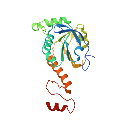
    Peroxiredoxins are antioxidant proteins primarily responsible for detoxification of hydroperoxides in cells. On exposure to various cellular stresses, peroxiredoxins can acquire chaperone activity, manifested as quaternary reorganization into a high molecular weight (HMW) form. Acidification, for example, causes dodecameric rings of human peroxiredoxin 3 (HsPrx3) to stack into long helical filaments. In this work, a 4.1-Å resolution structure of low-pH-instigated helical filaments was elucidated, showing a locally unfolded active site and partially folded C terminus. A 2.8-Å crystal structure of HsPrx3 was determined at pH 8.5 under reducing conditions, wherein dodecameric rings are arranged as a short stack, with symmetry similar to low-pH filaments. In contrast to previous observations, the crystal structure displays both a fully folded active site and ordered C terminus, suggesting that the HsPrx3 HMW form maintains catalytic activity. We propose a new role for the HMW form as a self-chaperoning assembly maintaining HsPrx3 function under stress.


  • Organizational Affiliation

    School of Biological Sciences, University of Auckland, Private Bag 92019, Auckland 1010, New Zealand; Biomolecular Interaction Centre and School of Biological Sciences, University of Canterbury, Private Bag 4800, Christchurch 8140, New Zealand.


Macromolecules
Find similar proteins by:  (by identity cutoff)  |  3D Structure
Entity ID: 1
MoleculeChains Sequence LengthOrganismDetailsImage
Thioredoxin-dependent peroxide reductase, mitochondrial
A, B, C, D, E
A, B, C, D, E, F, G, H, I
201Homo sapiensMutation(s): 0 
Gene Names: PRDX3AOP1
EC: 1.11.1.15
UniProt & NIH Common Fund Data Resources
Find proteins for P30048 (Homo sapiens)
Explore P30048 
Go to UniProtKB:  P30048
PHAROS:  P30048
GTEx:  ENSG00000165672 
Entity Groups  
Sequence Clusters30% Identity50% Identity70% Identity90% Identity95% Identity100% Identity
UniProt GroupP30048
Sequence Annotations
Expand
  • Reference Sequence
Experimental Data & Validation

Experimental Data

  • Method: X-RAY DIFFRACTION
  • Resolution: 2.80 Å
  • R-Value Free: 0.229 
  • R-Value Work: 0.183 
  • R-Value Observed: 0.185 
  • Space Group: I 2 2 2
Unit Cell:
Length ( Å )Angle ( ˚ )
a = 133.188α = 90
b = 168.73β = 90
c = 221.55γ = 90
Software Package:
Software NamePurpose
REFMACrefinement
MOSFLMdata reduction
Aimlessdata scaling
PHASERphasing

Structure Validation

View Full Validation Report


Entry History & Funding Information

Deposition Data


Funding OrganizationLocationGrant Number
RSNZNew ZealandRDF-UOA-1303

Revision History  (Full details and data files)

  • Version 1.0: 2016-05-25
    Type: Initial release
  • Version 1.1: 2016-06-15
    Changes: Database references
  • Version 1.2: 2016-07-20
    Changes: Database references
  • Version 1.3: 2023-09-27
    Changes: Data collection, Database references, Derived calculations, Refinement description