5LCF

VIM-2 metallo-beta-lactamase in complex with 3-oxo-2-phenylisoindoline-4-carboxylic acid (compound 30)


Experimental Data Snapshot

  • Method: X-RAY DIFFRACTION
  • Resolution: 1.86 Å
  • R-Value Free: 0.164 
  • R-Value Work: 0.127 
  • R-Value Observed: 0.130 

wwPDB Validation   3D Report Full Report


Ligand Structure Quality Assessment 


This is version 1.2 of the entry. See complete history


Literature

NMR-filtered virtual screening leads to non-metal chelating metallo-beta-lactamase inhibitors.

Li, G.B.Abboud, M.I.Brem, J.Someya, H.Lohans, C.T.Yang, S.Y.Spencer, J.Wareham, D.W.McDonough, M.A.Schofield, C.J.

(2017) Chem Sci 8: 928-937

  • DOI: https://doi.org/10.1039/c6sc04524c
  • Primary Citation of Related Structures:  
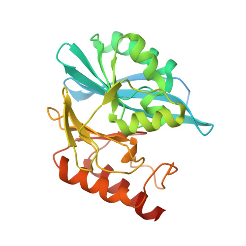
    5LCA, 5LCF, 5LCH, 5LE1, 5LM6

  • PubMed Abstract: 

    There are no clinically useful inhibitors of metallo-β-lactamases (MBLs), which are a growing problem because they hydrolyse almost all β-lactam antibacterials. Inhibition by most reported MBL inhibitors involves zinc ion chelation. A structure-based virtual screening approach combined with NMR filtering led to the identification of inhibitors of the clinically relevant Verona Integron-encoded MBL (VIM)-2. Crystallographic analyses reveal a new mode of MBL inhibition involving binding adjacent to the active site zinc ions, but which does not involve metal chelation. The results will aid efforts to develop new types of clinically useful inhibitors targeting MBLs/MBL-fold metallo-enzymes involved in antibacterial and anticancer drug resistance.


  • Organizational Affiliation

    Department of Chemistry , University of Oxford , 12 Mansfield Road , Oxford , OX1 3TA , UK . Email: christopher.schofield@chem.ox.ac.uk ; Email: michael.mcdonough@chem.ox.ac.uk.


Macromolecules
Find similar proteins by:  (by identity cutoff)  |  3D Structure
Entity ID: 1
MoleculeChains Sequence LengthOrganismDetailsImage
Metallo-beta-lactamase VIM-2266Pseudomonas aeruginosaMutation(s): 0 
Gene Names: blaVIM-2bla vim-2bla-VIM-2blasVIM-2blaVIM2blmVIM-2PAERUG_P32_London_17_VIM_2_10_11_06255
UniProt
Find proteins for Q9K2N0 (Pseudomonas aeruginosa)
Explore Q9K2N0 
Go to UniProtKB:  Q9K2N0
Entity Groups  
Sequence Clusters30% Identity50% Identity70% Identity90% Identity95% Identity100% Identity
UniProt GroupQ9K2N0
Sequence Annotations
Expand
  • Reference Sequence
Small Molecules
Binding Affinity Annotations 
IDSourceBinding Affinity
6TJ Binding MOAD:  5LCF IC50: 1.78e+4 (nM) from 1 assay(s)
Experimental Data & Validation

Experimental Data

  • Method: X-RAY DIFFRACTION
  • Resolution: 1.86 Å
  • R-Value Free: 0.164 
  • R-Value Work: 0.127 
  • R-Value Observed: 0.130 
  • Space Group: P 21 21 21
Unit Cell:
Length ( Å )Angle ( ˚ )
a = 40.46α = 90
b = 65.637β = 90
c = 70.721γ = 90
Software Package:
Software NamePurpose
SCALEPACKdata scaling
PHASERphasing
PHENIXrefinement
PDB_EXTRACTdata extraction
CrystalCleardata reduction
HKL-3000phasing

Structure Validation

View Full Validation Report


Ligand Structure Quality Assessment 


Entry History 

Deposition Data

Revision History  (Full details and data files)

  • Version 1.0: 2017-02-15
    Type: Initial release
  • Version 1.1: 2017-05-24
    Changes: Database references
  • Version 1.2: 2024-01-10
    Changes: Data collection, Database references, Derived calculations, Refinement description