5LFS

Lambda-[Ru(bpy)2(dppz)]2+ bound to brominated DNA

  • Classification: DNA
  • Organism(s): synthetic construct
  • Mutation(s): No 

  • Deposited: 2016-07-04 Released: 2017-02-15 
  • Deposition Author(s): Hall, J.P., Cardin, C.J.
  • Funding Organization(s): Biotechnology and Biological Sciences Research Council

Experimental Data Snapshot

  • Method: X-RAY DIFFRACTION
  • Resolution: 1.85 Å
  • R-Value Free: 0.192 
  • R-Value Work: 0.171 
  • R-Value Observed: 0.173 

wwPDB Validation   3D Report Full Report


Ligand Structure Quality Assessment 


This is version 1.2 of the entry. See complete history


Literature

Guanine Can Direct Binding Specificity of Ru-dipyridophenazine (dppz) Complexes to DNA through Steric Effects.

Hall, J.P.Gurung, S.P.Henle, J.Poidl, P.Andersson, J.Lincoln, P.Winter, G.Sorensen, T.Cardin, D.J.Brazier, J.A.Cardin, C.J.

(2017) Chemistry 23: 4981-4985

  • DOI: https://doi.org/10.1002/chem.201605508
  • Primary Citation of Related Structures:  
    5LFS, 5LFW, 5LFX

  • PubMed Abstract: 

    X-ray crystal structures of three Λ-[Ru(L) 2 dppz] 2+ complexes (dppz=dipyridophenazine; L=1,10-phenanthroline (phen), 2,2'-bipyridine (bpy)) bound to d((5BrC)GGC/GCCG) showed the compounds intercalated at a 5'-CG-3' step. The compounds bind through canted intercalation, with the binding angle determined by the guanine NH 2 group, in contrast to symmetrical intercalation previously observed at 5'-TA-3' sites. This result suggests that canted intercalation is preferred at 5'-CG-3' sites even though the site itself is symmetrical, and we hypothesise that symmetrical intercalation in a 5'-CG-3' step could give rise to a longer luminescence lifetime than canted intercalation.


  • Organizational Affiliation

    Department of Chemistry, University of Reading, Whiteknights, Reading, RG6 6AD, UK.


Macromolecules

Find similar nucleic acids by:  Sequence   |   3D Structure  

Entity ID: 1
MoleculeChains LengthOrganismImage
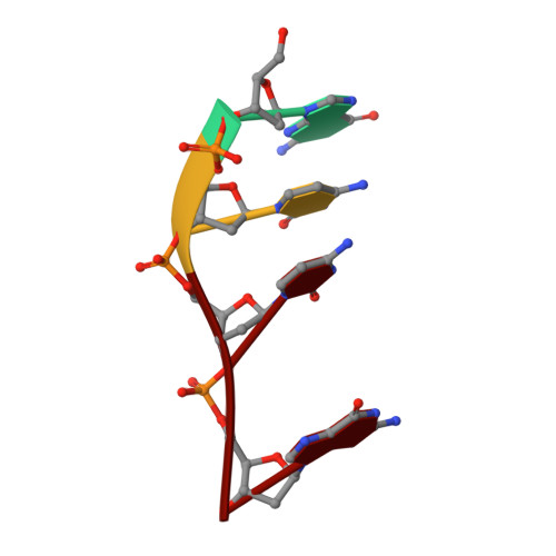
DNA (5'-D(*GP*CP*CP*G)-3')4synthetic construct
Sequence Annotations
Expand
  • Reference Sequence

Find similar nucleic acids by:  Sequence   |   3D Structure  

Entity ID: 2
MoleculeChains LengthOrganismImage
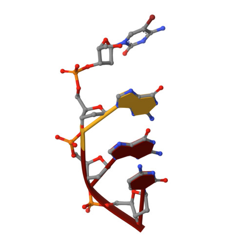
DNA (5'-D(*(CBR)P*GP*GP*C)-3')B [auth C]4synthetic construct
Sequence Annotations
Expand
  • Reference Sequence
Experimental Data & Validation

Experimental Data

  • Method: X-RAY DIFFRACTION
  • Resolution: 1.85 Å
  • R-Value Free: 0.192 
  • R-Value Work: 0.171 
  • R-Value Observed: 0.173 
  • Space Group: P 64 2 2
Unit Cell:
Length ( Å )Angle ( ˚ )
a = 66.9α = 90
b = 66.9β = 90
c = 36.02γ = 120
Software Package:
Software NamePurpose
REFMACrefinement
XDSdata reduction
Aimlessdata scaling
SHELXCDphasing
xia2data reduction

Structure Validation

View Full Validation Report


Ligand Structure Quality Assessment 


Entry History & Funding Information

Deposition Data


Funding OrganizationLocationGrant Number
Biotechnology and Biological Sciences Research CouncilUnited KingdomBB/K019279/1
Biotechnology and Biological Sciences Research CouncilUnited KingdomBB/M004635/1

Revision History  (Full details and data files)

  • Version 1.0: 2017-02-15
    Type: Initial release
  • Version 1.1: 2017-04-19
    Changes: Database references
  • Version 1.2: 2017-08-30
    Changes: Author supporting evidence