Structures and transport dynamics of a Campylobacter jejuni multidrug efflux pump.
Su, C.C., Yin, L., Kumar, N., Dai, L., Radhakrishnan, A., Bolla, J.R., Lei, H.T., Chou, T.H., Delmar, J.A., Rajashankar, K.R., Zhang, Q., Shin, Y.K., Yu, E.W.(2017) Nat Commun 8: 171-171
- PubMed: 28761097
- DOI: https://doi.org/10.1038/s41467-017-00217-z
- Primary Citation of Related Structures:
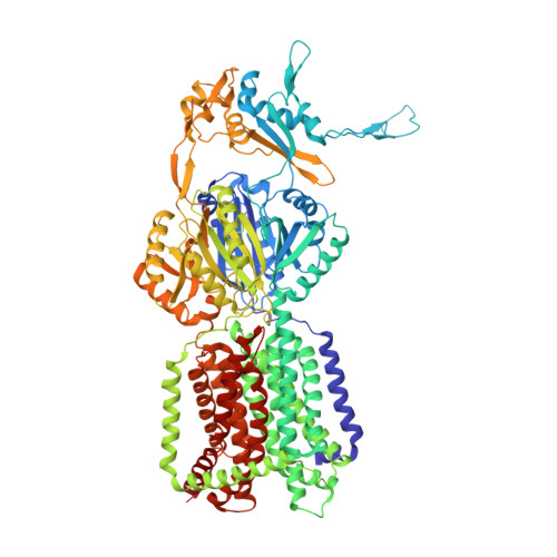
5LQ3, 5T0O - PubMed Abstract:
Resistance-nodulation-cell division efflux pumps are integral membrane proteins that catalyze the export of substrates across cell membranes. Within the hydrophobe-amphiphile efflux subfamily, these resistance-nodulation-cell division proteins largely form trimeric efflux pumps. The drug efflux process has been proposed to entail a synchronized motion between subunits of the trimer to advance the transport cycle, leading to the extrusion of drug molecules. Here we use X-ray crystallography and single-molecule fluorescence resonance energy transfer imaging to elucidate the structures and functional dynamics of the Campylobacter jejuni CmeB multidrug efflux pump. We find that the CmeB trimer displays a very unique conformation. A direct observation of transport dynamics in individual CmeB trimers embedded in membrane vesicles indicates that each CmeB subunit undergoes conformational transitions uncoordinated and independent of each other. On the basis of our findings and analyses, we propose a model for transport mechanism where CmeB protomers function independently within the trimer.Multidrug efflux pumps significantly contribute for bacteria resistance to antibiotics. Here the authors present the structure of Campylobacter jejuni CmeB pump combined with functional FRET assays to propose a transport mechanism where each CmeB protomers is functionally independent from the trimer.
Organizational Affiliation:
Department of Physics and Astronomy, Iowa State University, Ames, IA, 50011, USA.