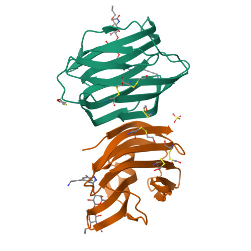
Galectin-1 in Complex with Ligand JB60
Grimm, C., Bechold, J.To be published.
Experimental Data Snapshot
Entity ID: 1 | |||||
|---|---|---|---|---|---|
| Molecule | Chains | Sequence Length | Organism | Details | Image |
| Galectin-1 | 133 | Homo sapiens | Mutation(s): 0 Gene Names: LGALS1 |  | |
UniProt & NIH Common Fund Data Resources | |||||
Find proteins for P09382 (Homo sapiens) Explore P09382 Go to UniProtKB: P09382 | |||||
PHAROS: P09382 GTEx: ENSG00000100097 | |||||
Entity Groups | |||||
| Sequence Clusters | 30% Identity50% Identity70% Identity90% Identity95% Identity100% Identity | ||||
| UniProt Group | P09382 | ||||
Sequence AnnotationsExpand | |||||
| |||||
| Ligands 3 Unique | |||||
|---|---|---|---|---|---|
| ID | Chains | Name / Formula / InChI Key | 2D Diagram | 3D Interactions | |
| WYD Query on WYD | D [auth A], F [auth B] | ~{N}-[(2~{R},3~{R},4~{R},5~{S},6~{R})-5-[(2~{S},3~{R},4~{S},5~{S},6~{R})-4-[[1-[[3-(3-azanylprop-1-ynyl)phenyl]methyl]-1,2,3-triazol-4-yl]methoxy]-6-(hydroxymethyl)-3,5-bis(oxidanyl)oxan-2-yl]oxy-6-(hydroxymethyl)-4-oxidanyl-2-propoxy-oxan-3-yl]ethanamide C30 H43 N5 O11 UZRLHTZEARMTAQ-JSXKIBNASA-N |  | ||
| SO4 Query on SO4 | E [auth B] | SULFATE ION O4 S QAOWNCQODCNURD-UHFFFAOYSA-L |  | ||
| BME Query on BME | C [auth A] | BETA-MERCAPTOETHANOL C2 H6 O S DGVVWUTYPXICAM-UHFFFAOYSA-N |  | ||
| Modified Residues 1 Unique | |||||
|---|---|---|---|---|---|
| ID | Chains | Type | Formula | 2D Diagram | Parent |
| CME Query on CME | A, B | L-PEPTIDE LINKING | C5 H11 N O3 S2 |  | CYS |
| Length ( Å ) | Angle ( ˚ ) |
|---|---|
| a = 43.26 | α = 90 |
| b = 58.25 | β = 90 |
| c = 111.25 | γ = 90 |
| Software Name | Purpose |
|---|---|
| PHENIX | refinement |
| PHASER | data processing |
| PDB_EXTRACT | data extraction |
| XDS | data reduction |
| PHASER | phasing |
| XDS | data scaling |
RCSB PDB is hosted by

RCSB PDB is a member of the