5NBE

[Ru(TAP)2(dppz-11-CN)]2+ bound to d(TCGGCGCCGA)2


Experimental Data Snapshot

  • Method: X-RAY DIFFRACTION
  • Resolution: 1.51 Å
  • R-Value Free: 0.190 
  • R-Value Work: 0.165 
  • R-Value Observed: 0.166 

wwPDB Validation   3D Report Full Report


Ligand Structure Quality Assessment 


This is version 2.1 of the entry. See complete history


Literature

X-ray Crystal Structures Show DNA Stacking Advantage of Terminal Nitrile Substitution in Ru-dppz Complexes.

McQuaid, K.Hall, J.P.Brazier, J.A.Cardin, D.J.Cardin, C.J.

(2018) Chemistry 24: 15859-15867

  • DOI: https://doi.org/10.1002/chem.201803021
  • Primary Citation of Related Structures:  
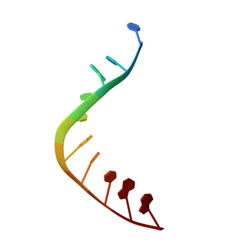
    5NBE, 6G8S, 6GLD, 6R6D

  • PubMed Abstract: 

    The new complexes [Ru(TAP) 2 (11-CN-dppz)] 2+ , [Ru(TAP) 2 (11-Br-dppz)] 2+ and [Ru(TAP) 2 (11,12-diCN-dppz)] 2+ are reported. The addition of nitrile substituents to the dppz ligand of the DNA photo-oxidising complex [Ru(TAP) 2 (dppz)] 2+ promote π-stacking interactions and ordered binding to DNA, as shown by X-ray crystallography. The structure of Λ-[Ru(TAP) 2 (11-CN-dppz)] 2+ with the DNA duplex d(TCGGCGCCGA) 2 shows, for the first time with this class of complex, a closed intercalation cavity with an AT base pair at the terminus. The structure obtained is compared to that formed with the 11-Br and 11,12-dinitrile derivatives, highlighting the stabilization of syn guanine by this enantiomer when the terminal base pair is GC. In contrast the AT base pair has the normal Watson-Crick orientation, highlighting the difference in charge distribution between the two purine bases and the complementarity of the dppz-purine interaction. The asymmetry of the cavity highlights the importance of the purine-dppz-purine stacking interaction.


  • Organizational Affiliation

    Department of Chemistry, University of Reading, Whiteknights, Reading, RG6 6AD, UK.


Macromolecules

Find similar nucleic acids by:  Sequence   |   3D Structure  

Entity ID: 1
MoleculeChains LengthOrganismImage
DNA (5'-D(*TP*CP*GP*GP*CP*GP*CP*CP*GP*A)-3')10synthetic construct
Sequence Annotations
Expand
  • Reference Sequence
Small Molecules
Ligands 2 Unique
IDChains Name / Formula / InChI Key2D Diagram3D Interactions
KSB
Query on KSB

Download Ideal Coordinates CCD File 
B [auth A]lambda-[Ru(tap2-dppz-CN)]2+
C39 H21 N13 Ru
HNFPSJMHTKCUIN-UHFFFAOYSA-N
BA
Query on BA

Download Ideal Coordinates CCD File 
C [auth A]BARIUM ION
Ba
XDFCIPNJCBUZJN-UHFFFAOYSA-N
Experimental Data & Validation

Experimental Data

  • Method: X-RAY DIFFRACTION
  • Resolution: 1.51 Å
  • R-Value Free: 0.190 
  • R-Value Work: 0.165 
  • R-Value Observed: 0.166 
  • Space Group: P 43 21 2
Unit Cell:
Length ( Å )Angle ( ˚ )
a = 47.88α = 90
b = 47.88β = 90
c = 29.14γ = 90
Software Package:
Software NamePurpose
REFMACrefinement
XDSdata reduction
XSCALEdata scaling
SHELXDEphasing
xia2data reduction
Cootmodel building

Structure Validation

View Full Validation Report


Ligand Structure Quality Assessment 


Entry History & Funding Information

Deposition Data


Funding OrganizationLocationGrant Number
Biotechnology and Biological Sciences Research CouncilUnited KingdomBB/K019279/1
Biotechnology and Biological Sciences Research CouncilUnited KingdomBB/M004635/1

Revision History  (Full details and data files)

  • Version 1.0: 2018-03-21
    Type: Initial release
  • Version 1.1: 2019-08-28
    Changes: Data collection, Database references
  • Version 2.0: 2023-12-27
    Changes: Atomic model, Data collection, Database references, Derived calculations, Non-polymer description, Structure summary
  • Version 2.1: 2024-01-10
    Changes: Derived calculations, Structure summary