5NYJ

Anbu from Hyphomicrobium sp. strain MC1 - SG: C2


Experimental Data Snapshot

  • Method: X-RAY DIFFRACTION
  • Resolution: 3.20 Å
  • R-Value Free: 0.279 
  • R-Value Work: 0.235 
  • R-Value Observed: 0.237 

wwPDB Validation   3D Report Full Report


This is version 1.3 of the entry. See complete history


Literature

On the Trails of the Proteasome Fold: Structural and Functional Analysis of the Ancestral beta-Subunit Protein Anbu.

Vielberg, M.T.Bauer, V.C.Groll, M.

(2018) J Mol Biol 430: 628-640

  • DOI: https://doi.org/10.1016/j.jmb.2018.01.004
  • Primary Citation of Related Structures:  
    5NYF, 5NYG, 5NYJ, 5NYP, 5NYQ, 5NYR

  • PubMed Abstract: 

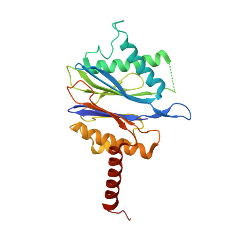
    The 20S proteasome is a key player in eukaryotic and archaeal protein degradation, but its progenitor in eubacteria is unknown. Recently, the ancestral β-subunit protein (Anbu) was predicted to be the evolutionary precursor of the proteasome. We crystallized Anbu from Hyphomicrobium sp. strain MC1 in four different space groups and solved the structures by SAD-phasing and Patterson search calculation techniques. Our data reveal that Anbu adopts the classical fold of Ntn-hydrolases, but its oligomeric state differs from that of barrel-shaped proteases. In contrast to their typical architecture, the Anbu protomer is a tightly interacting dimer that can assemble into a helical superstructure. Although Anbu features a catalytic triad of Thr1O γ , Asp17O δ1 and Lys32N ε , it is unable to hydrolyze standard protease substrates. The lack of activity might be caused by the incapacity of Thr1NH 2 to function as a Brønsted acid during substrate cleavage due to its missing activation via hydrogen bonding. Altogether, we demonstrate that the topology of the proteasomal fold is conserved in Anbu, but whether it acts as a protease still needs to be clarified.


  • Organizational Affiliation

    Center for Integrated Protein Science Munich at the Department Chemie, Lehrstuhl für Biochemie, Technische Universität München, Garching, Germany.


Macromolecules
Find similar proteins by:  (by identity cutoff)  |  3D Structure
Entity ID: 1
MoleculeChains Sequence LengthOrganismDetailsImage
Anbu
A, B, C, D, E
A, B, C, D, E, F, G, H, I, J, K, L, M, N, O, P
244Hyphomicrobium sp. MC1Mutation(s): 0 
Gene Names: HYPMC_4374
UniProt
Find proteins for F8JB59 (Hyphomicrobium sp. (strain MC1))
Explore F8JB59 
Go to UniProtKB:  F8JB59
Entity Groups  
Sequence Clusters30% Identity50% Identity70% Identity90% Identity95% Identity100% Identity
UniProt GroupF8JB59
Sequence Annotations
Expand
  • Reference Sequence
Experimental Data & Validation

Experimental Data

  • Method: X-RAY DIFFRACTION
  • Resolution: 3.20 Å
  • R-Value Free: 0.279 
  • R-Value Work: 0.235 
  • R-Value Observed: 0.237 
  • Space Group: C 1 2 1
Unit Cell:
Length ( Å )Angle ( ˚ )
a = 240.22α = 90
b = 242.15β = 90.77
c = 107.32γ = 90
Software Package:
Software NamePurpose
REFMACrefinement
XDSdata reduction
XSCALEdata scaling
PHASERphasing

Structure Validation

View Full Validation Report


Entry History & Funding Information

Deposition Data


Funding OrganizationLocationGrant Number
German Research FoundationGermanySFB1035

Revision History  (Full details and data files)

  • Version 1.0: 2017-05-24
    Type: Initial release
  • Version 1.1: 2018-02-28
    Changes: Author supporting evidence, Database references
  • Version 1.2: 2018-03-07
    Changes: Database references
  • Version 1.3: 2024-01-17
    Changes: Data collection, Database references, Refinement description