5O86

Mutant of claas II CPD photolyase from Methanosarcina mazei - W388F


Experimental Data Snapshot

  • Method: X-RAY DIFFRACTION
  • Resolution: 1.69 Å
  • R-Value Free: 0.243 
  • R-Value Work: 0.205 
  • R-Value Observed: 0.206 

wwPDB Validation   3D Report Full Report


Ligand Structure Quality Assessment 


This is version 1.4 of the entry. See complete history


Literature

Nicotinamide Adenine Dinucleotides Arrest Photoreduction of Class II DNA Photolyases in FADH ̇ State.

Ignatz, E.Geisselbrecht, Y.Kiontke, S.Essen, L.O.

(2018) Photochem Photobiol 94: 81-87

  • DOI: https://doi.org/10.1111/php.12834
  • Primary Citation of Related Structures:  
    5O86, 5O8D, 5O8E

  • PubMed Abstract: 

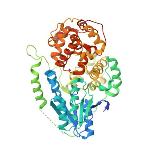
    All light-sensitive members of the photolyase/cryptochrome family rely on FAD as catalytic cofactor. Its activity is regulated by photoreduction, a light-triggered electron transfer process from a conserved tryptophan triad to the flavin. The stability of the reduced flavin depends on available external electron donors and oxygen. In this study, we show for the class II photolyase of Methanosarcina mazei, MmCPDII, that it utilizes physiologically relevant redox cofactors NADH and NADPH for the formation of the semiquinoid FAD in a light-dependent reaction. Using redox-inert variants MmCPDII/W388F and MmCPDII/W360F, we demonstrate that photoreduction by NADH and NADPH requires the class II-specific tryptophan cascade of MmCPDII. Finally, we confirmed that mutations in the tryptophan cascade can be introduced without any substantial structural disturbances by analyzing crystal structures of MmCPDII/W388F, MmCPDII/W360F and MmCPDII/Y345F.


  • Organizational Affiliation

    Department of Chemistry, LOEWE Center for Synthetic Microbiology, Philipps University, Marburg, Germany.


Macromolecules
Find similar proteins by:  (by identity cutoff)  |  3D Structure
Entity ID: 1
MoleculeChains Sequence LengthOrganismDetailsImage
Deoxyribodipyrimidine photolyase484Methanosarcina mazei Go1Mutation(s): 2 
Gene Names: MM_0852
EC: 4.1.99.3
UniProt
Find proteins for Q8PYK9 (Methanosarcina mazei (strain ATCC BAA-159 / DSM 3647 / Goe1 / Go1 / JCM 11833 / OCM 88))
Explore Q8PYK9 
Go to UniProtKB:  Q8PYK9
Entity Groups  
Sequence Clusters30% Identity50% Identity70% Identity90% Identity95% Identity100% Identity
UniProt GroupQ8PYK9
Sequence Annotations
Expand
  • Reference Sequence
Small Molecules
Ligands 3 Unique
IDChains Name / Formula / InChI Key2D Diagram3D Interactions
FAD
Query on FAD

Download Ideal Coordinates CCD File 
B [auth A]FLAVIN-ADENINE DINUCLEOTIDE
C27 H33 N9 O15 P2
VWWQXMAJTJZDQX-UYBVJOGSSA-N
SO4
Query on SO4

Download Ideal Coordinates CCD File 
C [auth A]
D [auth A]
E [auth A]
F [auth A]
G [auth A]
C [auth A],
D [auth A],
E [auth A],
F [auth A],
G [auth A],
H [auth A],
I [auth A],
J [auth A],
K [auth A]
SULFATE ION
O4 S
QAOWNCQODCNURD-UHFFFAOYSA-L
GOL
Query on GOL

Download Ideal Coordinates CCD File 
L [auth A],
M [auth A]
GLYCEROL
C3 H8 O3
PEDCQBHIVMGVHV-UHFFFAOYSA-N
Experimental Data & Validation

Experimental Data

  • Method: X-RAY DIFFRACTION
  • Resolution: 1.69 Å
  • R-Value Free: 0.243 
  • R-Value Work: 0.205 
  • R-Value Observed: 0.206 
  • Space Group: P 43 21 2
Unit Cell:
Length ( Å )Angle ( ˚ )
a = 69.72α = 90
b = 69.72β = 90
c = 243.85γ = 90
Software Package:
Software NamePurpose
PHENIXrefinement
REFMACrefinement

Structure Validation

View Full Validation Report


Ligand Structure Quality Assessment 


Entry History 

Deposition Data

Revision History  (Full details and data files)

  • Version 1.0: 2017-09-13
    Type: Initial release
  • Version 1.1: 2017-11-08
    Changes: Database references
  • Version 1.2: 2017-11-22
    Changes: Database references
  • Version 1.3: 2018-01-24
    Changes: Database references
  • Version 1.4: 2019-10-16
    Changes: Data collection