5U7N

CRYSTAL STRUCTURE OF A CHIMERIC CUA DOMAIN (SUBUNIT II) OF CYTOCHROME BA3 FROM THERMUS THERMOPHILUS WITH THE AMICYANIN LOOP


Experimental Data Snapshot

  • Method: X-RAY DIFFRACTION
  • Resolution: 2.30 Å
  • R-Value Free: 0.226 
  • R-Value Work: 0.176 
  • R-Value Observed: 0.178 

wwPDB Validation   3D Report Full Report


This is version 1.3 of the entry. See complete history


Literature

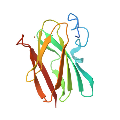
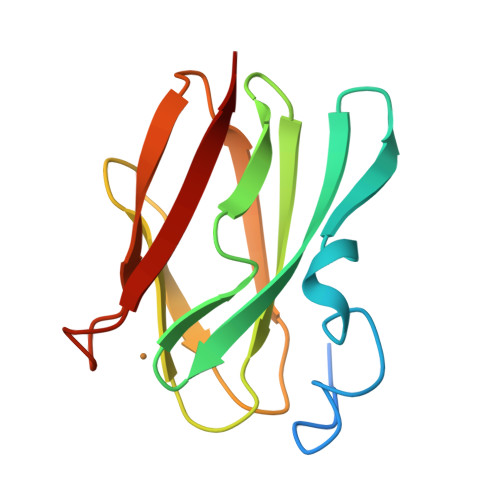
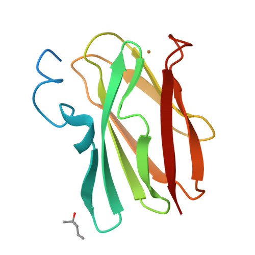
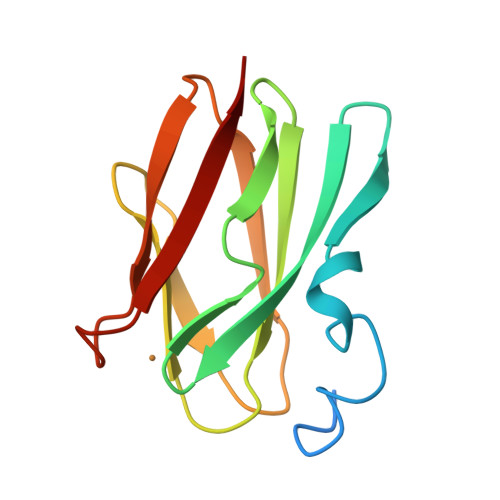
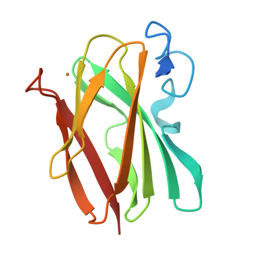
Engineering a bifunctional copper site in the cupredoxin fold by loop-directed mutagenesis.

Espinoza-Cara, A.Zitare, U.Alvarez-Paggi, D.Klinke, S.Otero, L.H.Murgida, D.H.Vila, A.J.

(2018) Chem Sci 9: 6692-6702

  • DOI: https://doi.org/10.1039/c8sc01444b
  • Primary Citation of Related Structures:  
    5U7N

  • PubMed Abstract: 

    Copper sites in proteins are designed to perform either electron transfer or redox catalysis. Type 1 and Cu A sites are electron transfer hubs bound to a rigid protein fold that prevents binding of exogenous ligands and side reactions. Here we report the engineering of two Type 1 sites by loop-directed mutagenesis within a Cu A scaffold with unique electronic structures and functional features. A copper-thioether axial bond shorter than the copper-thiolate bond is responsible for the electronic structure features, in contrast to all other natural or chimeric sites where the copper thiolate bond is short. These sites display highly unusual features, such as: (1) a high reduction potential despite a strong interaction with the axial ligand, which we attribute to changes in the hydrogen bond network and (2) the ability to bind exogenous ligands such as imidazole and azide. This strategy widens the possibility of using natural protein scaffolds with functional features not present in nature.


  • Organizational Affiliation

    Instituto de Biología Molecular y Celular de Rosario (IBR, CONICET-UNR) , Rosario , Argentina . Email: vila@ibr-conicet.gov.ar.


Macromolecules
Find similar proteins by:  (by identity cutoff)  |  3D Structure
Entity ID: 1
MoleculeChains Sequence LengthOrganismDetailsImage
Cytochrome c oxidase subunit 2
A, B, C, D, E
A, B, C, D, E, F, G, H
124Thermus thermophilusMutation(s): 0 
Gene Names: cbaBctaC
EC: 1.9.3.1
UniProt
Find proteins for Q5SJ80 (Thermus thermophilus (strain ATCC 27634 / DSM 579 / HB8))
Explore Q5SJ80 
Go to UniProtKB:  Q5SJ80
Entity Groups  
Sequence Clusters30% Identity50% Identity70% Identity90% Identity95% Identity100% Identity
UniProt GroupQ5SJ80
Sequence Annotations
Expand
  • Reference Sequence
Small Molecules
Ligands 2 Unique
IDChains Name / Formula / InChI Key2D Diagram3D Interactions
MPD
Query on MPD

Download Ideal Coordinates CCD File 
L [auth C],
P [auth F]
(4S)-2-METHYL-2,4-PENTANEDIOL
C6 H14 O2
SVTBMSDMJJWYQN-YFKPBYRVSA-N
CU
Query on CU

Download Ideal Coordinates CCD File 
I [auth A]
J [auth B]
K [auth C]
M [auth D]
N [auth E]
I [auth A],
J [auth B],
K [auth C],
M [auth D],
N [auth E],
O [auth F],
Q [auth G],
R [auth H]
COPPER (II) ION
Cu
JPVYNHNXODAKFH-UHFFFAOYSA-N
Experimental Data & Validation

Experimental Data

  • Method: X-RAY DIFFRACTION
  • Resolution: 2.30 Å
  • R-Value Free: 0.226 
  • R-Value Work: 0.176 
  • R-Value Observed: 0.178 
  • Space Group: P 21 21 21
Unit Cell:
Length ( Å )Angle ( ˚ )
a = 94.352α = 90
b = 100.701β = 90
c = 101.103γ = 90
Software Package:
Software NamePurpose
Aimlessdata scaling
PHASERphasing
BUSTERrefinement
PDB_EXTRACTdata extraction
XDSdata reduction

Structure Validation

View Full Validation Report


Entry History 

Deposition Data

Revision History  (Full details and data files)

  • Version 1.0: 2017-11-22
    Type: Initial release
  • Version 1.1: 2017-11-29
    Changes: Database references, Structure summary
  • Version 1.2: 2018-12-05
    Changes: Data collection, Database references
  • Version 1.3: 2023-10-04
    Changes: Data collection, Database references, Refinement description