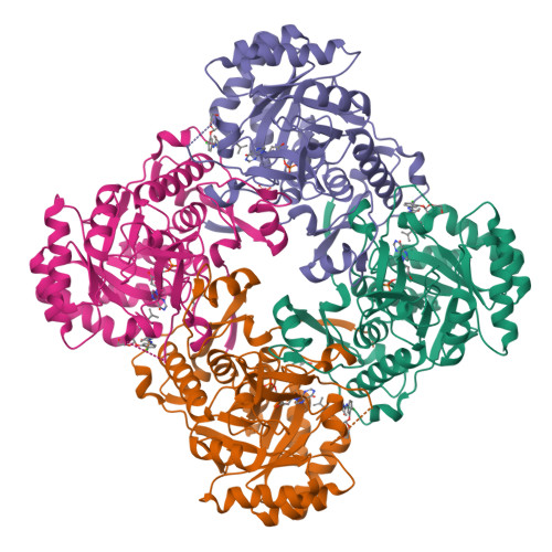
5UXE | pdb_00005uxe

Crystal Structure of Inosine 5'-monophosphate Dehydrogenase from Clostridium perfringens Complexed with IMP and P178


Experimental Data Snapshot

  • Method: X-RAY DIFFRACTION
  • Resolution: 2.10 Å
  • R-Value Free: 
    0.223 (Depositor), 0.220 (DCC) 
  • R-Value Work: 
    0.173 (Depositor), 0.170 (DCC) 
  • R-Value Observed: 
    0.176 (Depositor) 

Starting Model: experimental
View more details

wwPDB Validation   3D Report Full Report


Ligand Structure Quality Assessment 

Created with Raphaël 2.3.0Worse 01 BetterLigand structure goodness of fit to experimental dataBest fitted 8LAClick on this verticalbar to view detailsBest fitted IMPClick on this verticalbar to view details

This is version 1.1 of the entry. See complete history


Literature

Crystal Structure of Inosine 5'-monophosphate Dehydrogenase from Clostridium perfringens Complexed with IMP and P178

Maltseva, N.Kim, Y.Mulligan, R.Makowska-Grzyska, M.Gu, M.Gollapalli, D.R.Hedstrom, L.Joachimiak, A.Anderson, W.F.Center for Structural Genomics of Infectious Diseases (CSGID)

To be published.

Macromolecules
Find similar proteins by:  (by identity cutoff)  |  3D Structure
Entity ID: 1
MoleculeChains Sequence LengthOrganismDetailsImage
Inosine-5'-monophosphate dehydrogenase
A, B, C, D
363Clostridium perfringensMutation(s): 0 
Gene Names: guaB_2guaBERS852446_02285JFP838_13815
EC: 1.1.1.205
UniProt
Find proteins for A0A127ELD1 (Clostridium perfringens)
Explore A0A127ELD1 
Go to UniProtKB:  A0A127ELD1
Entity Groups  
Sequence Clusters30% Identity50% Identity70% Identity90% Identity95% Identity100% Identity
UniProt GroupA0A127ELD1
Sequence Annotations
Expand
  • Reference Sequence
Small Molecules
Ligands 6 Unique
IDChains Name / Formula / InChI Key2D Diagram3D Interactions
8LA
Query on 8LA

Download Ideal Coordinates CCD File 
F [auth A],
I [auth B],
N [auth C],
R [auth D]
N-[4-chloro-3-(alpha-D-ribofuranosyloxy)phenyl]-N'-{2-[3-(prop-1-en-2-yl)phenyl]propan-2-yl}urea
C24 H29 Cl N2 O6
DGCHEIBDGDMRPM-YSFYHYPLSA-N
IMP
Query on IMP

Download Ideal Coordinates CCD File 
E [auth A],
H [auth B],
M [auth C],
Q [auth D]
INOSINIC ACID
C10 H13 N4 O8 P
GRSZFWQUAKGDAV-KQYNXXCUSA-N
MPD
Query on MPD

Download Ideal Coordinates CCD File 
J [auth B],
S [auth D]
(4S)-2-METHYL-2,4-PENTANEDIOL
C6 H14 O2
SVTBMSDMJJWYQN-YFKPBYRVSA-N
SO4
Query on SO4

Download Ideal Coordinates CCD File 
O [auth C]SULFATE ION
O4 S
QAOWNCQODCNURD-UHFFFAOYSA-L
FMT
Query on FMT

Download Ideal Coordinates CCD File 
L [auth C]FORMIC ACID
C H2 O2
BDAGIHXWWSANSR-UHFFFAOYSA-N
K
Query on K

Download Ideal Coordinates CCD File 
G [auth A],
K [auth B],
P [auth D]
POTASSIUM ION
K
NPYPAHLBTDXSSS-UHFFFAOYSA-N
Experimental Data & Validation

Experimental Data

  • Method: X-RAY DIFFRACTION
  • Resolution: 2.10 Å
  • R-Value Free:  0.223 (Depositor), 0.220 (DCC) 
  • R-Value Work:  0.173 (Depositor), 0.170 (DCC) 
  • R-Value Observed: 0.176 (Depositor) 
Space Group: C 1 2 1
Unit Cell:
Length ( Å )Angle ( ˚ )
a = 127.937α = 90
b = 119.101β = 102.86
c = 96.971γ = 90
Software Package:
Software NamePurpose
PHENIXrefinement
HKL-3000data reduction
HKL-3000data scaling
HKL-3000phasing

Structure Validation

View Full Validation Report


Ligand Structure Quality Assessment 

Created with Raphaël 2.3.0Worse 01 BetterLigand structure goodness of fit to experimental dataBest fitted 8LAClick on this verticalbar to view detailsBest fitted IMPClick on this verticalbar to view details

Entry History 

Revision History  (Full details and data files)

  • Version 1.0: 2017-03-08
    Type: Initial release
  • Version 1.1: 2023-10-04
    Changes: Data collection, Database references, Derived calculations, Refinement description