8N1
Query on 8N1
Download Ideal Coordinates CCD File
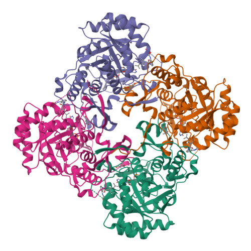
| F [auth A],
K [auth B],
O [auth C],
S [auth D] | N-{2-chloro-5-[({2-[3-(prop-1-en-2-yl)phenyl]propan-2-yl}carbamoyl)amino]phenyl}-beta-D-xylofuranosylamine
C24 H30 Cl N3 O5
RGENKIJBZZZCQP-CIAFKFPVSA-N |  | |
IMP
Query on IMP
Download Ideal Coordinates CCD File
| E [auth A],
J [auth B],
N [auth C],
R [auth D] | INOSINIC ACID
C10 H13 N4 O8 P
GRSZFWQUAKGDAV-KQYNXXCUSA-N |  | |
MRD
Query on MRD
Download Ideal Coordinates CCD File
| P [auth C] | (4R)-2-METHYLPENTANE-2,4-DIOL
C6 H14 O2
SVTBMSDMJJWYQN-RXMQYKEDSA-N |  | |
MPD
Query on MPD
Download Ideal Coordinates CCD File
| G [auth A],
L [auth B],
T [auth D] | (4S)-2-METHYL-2,4-PENTANEDIOL
C6 H14 O2
SVTBMSDMJJWYQN-YFKPBYRVSA-N |  | |
ACY
Query on ACY
Download Ideal Coordinates CCD File
| H [auth A],
Q [auth C],
U [auth D] | ACETIC ACID
C2 H4 O2
QTBSBXVTEAMEQO-UHFFFAOYSA-N |  | |
FMT
Query on FMT
Download Ideal Coordinates CCD File
| I [auth A],
M [auth B] | FORMIC ACID
C H2 O2
BDAGIHXWWSANSR-UHFFFAOYSA-N |  | |