5X58

Prefusion structure of SARS-CoV spike glycoprotein, conformation 1

  • Classification: VIRAL PROTEIN
  • Organism(s): SARS coronavirus BJ01
  • Expression System: Spodoptera frugiperda
  • Mutation(s): No 

  • Deposited: 2017-02-15 Released: 2017-05-03 
  • Deposition Author(s): Yuan, Y., Cao, D., Zhang, Y., Ma, J., Qi, J., Wang, Q., Lu, G., Wu, Y., Yan, J., Shi, Y., Zhang, X., Gao, G.F.
  • Funding Organization(s): Strategic Priority Research Program of the Chinese Academy of Sciences, China Ministry of Science and Technology (MOST) National 973 Project, the National Key Research and Development Program of China, the China National Grand S&T Special Project, the Natural Science Foundation of China, the Excellent Young Scientist Program from the NSFC, Excellent Young Scientist Program of the Chinese Academy of Sciences and the Youth Innovation Promotion Association CAS, National Thousand (Young) Talents Program, leading principal investigator of the NSFC Innovative Research Group

Experimental Data Snapshot

  • Method: ELECTRON MICROSCOPY
  • Resolution: 3.20 Å
  • Aggregation State: PARTICLE 
  • Reconstruction Method: SINGLE PARTICLE 

wwPDB Validation   3D Report Full Report


This is version 1.1 of the entry. See complete history


Literature

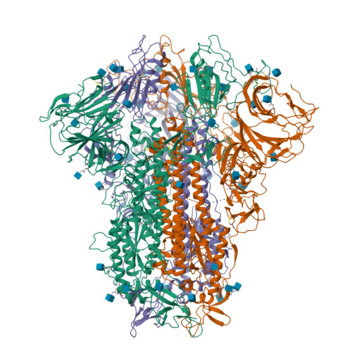
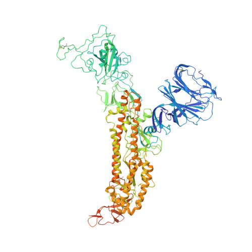
Cryo-EM structures of MERS-CoV and SARS-CoV spike glycoproteins reveal the dynamic receptor binding domains

Yuan, Y.Cao, D.Zhang, Y.Ma, J.Qi, J.Wang, Q.Lu, G.Wu, Y.Yan, J.Shi, Y.Zhang, X.Gao, G.F.

(2017) Nat Commun 8: 15092-15092

  • DOI: https://doi.org/10.1038/ncomms15092
  • Primary Citation of Related Structures:  
    5X4R, 5X4S, 5X58, 5X59, 5X5B, 5X5C, 5X5F

  • PubMed Abstract: 

    The envelope spike (S) proteins of MERS-CoV and SARS-CoV determine the virus host tropism and entry into host cells, and constitute a promising target for the development of prophylactics and therapeutics. Here, we present high-resolution structures of the trimeric MERS-CoV and SARS-CoV S proteins in its pre-fusion conformation by single particle cryo-electron microscopy. The overall structures resemble that from other coronaviruses including HKU1, MHV and NL63 reported recently, with the exception of the receptor binding domain (RBD). We captured two states of the RBD with receptor binding region either buried (lying state) or exposed (standing state), demonstrating an inherently flexible RBD readily recognized by the receptor. Further sequence conservation analysis of six human-infecting coronaviruses revealed that the fusion peptide, HR1 region and the central helix are potential targets for eliciting broadly neutralizing antibodies.


  • Organizational Affiliation

    School of Life Sciences, University of Science and Technology of China, Hefei, Anhui 230026, China.


Macromolecules
Find similar proteins by:  (by identity cutoff)  |  3D Structure
Entity ID: 1
MoleculeChains Sequence LengthOrganismDetailsImage
Spike glycoprotein
A, B, C
1,228SARS coronavirus BJ01Mutation(s): 0 
UniProt
Find proteins for P59594 (Severe acute respiratory syndrome coronavirus)
Explore P59594 
Go to UniProtKB:  P59594
Entity Groups  
Sequence Clusters30% Identity50% Identity70% Identity90% Identity95% Identity100% Identity
UniProt GroupP59594
Sequence Annotations
Expand
  • Reference Sequence
Small Molecules
Ligands 1 Unique
IDChains Name / Formula / InChI Key2D Diagram3D Interactions
NAG
Query on NAG

Download Ideal Coordinates CCD File 
AA [auth B]
BA [auth B]
CA [auth B]
D [auth A]
DA [auth B]
AA [auth B],
BA [auth B],
CA [auth B],
D [auth A],
DA [auth B],
E [auth A],
EA [auth B],
F [auth A],
FA [auth C],
G [auth A],
GA [auth C],
H [auth A],
HA [auth C],
I [auth A],
IA [auth C],
J [auth A],
JA [auth C],
K [auth A],
KA [auth C],
L [auth A],
LA [auth C],
M [auth A],
MA [auth C],
N [auth A],
NA [auth C],
O [auth A],
OA [auth C],
P [auth A],
PA [auth C],
Q [auth A],
QA [auth C],
R [auth B],
RA [auth C],
S [auth B],
SA [auth C],
T [auth B],
U [auth B],
V [auth B],
W [auth B],
X [auth B],
Y [auth B],
Z [auth B]
2-acetamido-2-deoxy-beta-D-glucopyranose
C8 H15 N O6
OVRNDRQMDRJTHS-FMDGEEDCSA-N
Experimental Data & Validation

Experimental Data

  • Method: ELECTRON MICROSCOPY
  • Resolution: 3.20 Å
  • Aggregation State: PARTICLE 
  • Reconstruction Method: SINGLE PARTICLE 

Structure Validation

View Full Validation Report


Entry History & Funding Information

Deposition Data


Funding OrganizationLocationGrant Number
Strategic Priority Research Program of the Chinese Academy of SciencesChinaXDB08020100
China Ministry of Science and Technology (MOST) National 973 ProjectChina2013CB531502
China Ministry of Science and Technology (MOST) National 973 ProjectChina2014CB542503
the National Key Research and Development Program of ChinaChina2016YFD0500300
the China National Grand S&T Special ProjectChina2014ZX10004-001-006
the Natural Science Foundation of ChinaChina31570874
the Natural Science Foundation of ChinaChina81461168030
the Excellent Young Scientist Program from the NSFCChina81622031
Excellent Young Scientist Program of the Chinese Academy of Sciences and the Youth Innovation Promotion Association CASChina2015078
National Thousand (Young) Talents ProgramChina--
leading principal investigator of the NSFC Innovative Research GroupChina81321063

Revision History  (Full details and data files)

  • Version 1.0: 2017-05-03
    Type: Initial release
  • Version 1.1: 2020-07-29
    Type: Remediation
    Reason: Carbohydrate remediation
    Changes: Data collection, Derived calculations, Structure summary