5XZJ

Crystal structure of the Zn-directed tetramer of the engineered cyt cb562 variant, C96T/AB5


Experimental Data Snapshot

  • Method: X-RAY DIFFRACTION
  • Resolution: 1.98 Å
  • R-Value Free: 0.217 
  • R-Value Work: 0.177 
  • R-Value Observed: 0.179 

wwPDB Validation   3D Report Full Report


Ligand Structure Quality Assessment 


This is version 2.1 of the entry. See complete history


Literature

Importance of Scaffold Flexibility/Rigidity in the Design and Directed Evolution of Artificial Metallo-beta-lactamases.

Song, W.J.Yu, J.Tezcan, F.A.

(2017) J Am Chem Soc 139: 16772-16779

  • DOI: https://doi.org/10.1021/jacs.7b08981
  • Primary Citation of Related Structures:  
    5XZI, 5XZJ

  • PubMed Abstract: 

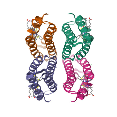
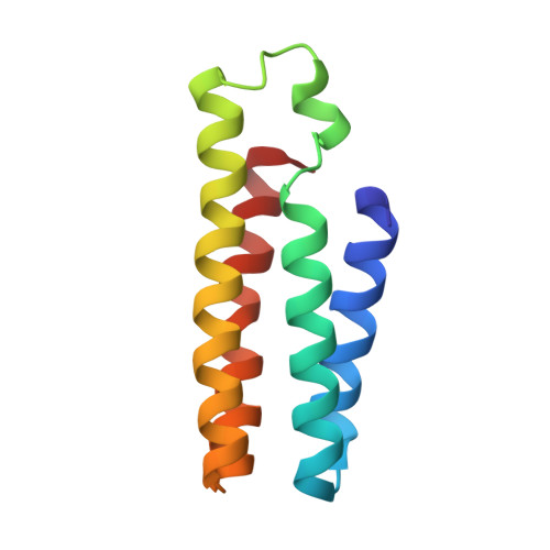
    We describe the design and evolution of catalytic hydrolase activity on a supramolecular protein scaffold, Zn 4 : C96 RIDC1 4 , which was constructed from cytochrome cb 562 building blocks via a metal-templating strategy. Previously, we reported that Zn 4 : C96 RIDC1 4 could be tailored with tripodal (His/His/Glu), unsaturated Zn coordination motifs in its interfaces to generate a variant termed Zn 8 : A104 AB3 4 , which in turn displayed catalytic activity for the hydrolysis of activated esters and β-lactam antibiotics. Zn 8 : A104 AB3 4 was subsequently subjected to directed evolution via an in vivo selection strategy, leading to a variant Zn 8 : A104/G57 AB3 4 which displayed enzyme-like Michaelis-Menten behavior for ampicillin hydrolysis. A criterion for the evolutionary utility or designability of a new protein structure is its ability to accommodate different active sites. With this in mind, we examined whether Zn 4 : C96 RIDC1 4 could be tailored with alternative Zn coordination sites that could similarly display evolvable catalytic activities. We report here a detailed structural and functional characterization of new variant Zn 8 :AB5 4 , which houses similar, unsaturated Zn coordination sites to those in Zn 8 : A104/G57 AB3 4 , but in completely different microenvironments. Zn 8 :AB5 4 displays Michaelis-Menten behavior for ampicillin hydrolysis without any optimization. Yet, the subsequent directed evolution of Zn 8 :AB5 4 revealed limited catalytic improvement, which we ascribed to the local protein rigidity surrounding the Zn centers and the lack of evolvable loop structures nearby. The relaxation of local rigidity via the elimination of adjacent disulfide linkages led to a considerable structural transformation with a concomitant improvement in β-lactamase activity. Our findings reaffirm previous observations that the delicate balance between protein flexibility and stability is crucial for enzyme design and evolution.


  • Organizational Affiliation

    Department of Chemistry and Biochemistry, University of California, San Diego , La Jolla, California 92093-0356, United States.


Macromolecules
Find similar proteins by:  (by identity cutoff)  |  3D Structure
Entity ID: 1
MoleculeChains Sequence LengthOrganismDetailsImage
Soluble cytochrome b562
A, B, C, D
106Escherichia coliMutation(s): 15 
Gene Names: cybC
UniProt
Find proteins for P0ABE7 (Escherichia coli)
Explore P0ABE7 
Go to UniProtKB:  P0ABE7
Entity Groups  
Sequence Clusters30% Identity50% Identity70% Identity90% Identity95% Identity100% Identity
UniProt GroupP0ABE7
Sequence Annotations
Expand
  • Reference Sequence
Small Molecules
Ligands 3 Unique
IDChains Name / Formula / InChI Key2D Diagram3D Interactions
HEC
Query on HEC

Download Ideal Coordinates CCD File 
E [auth A],
K [auth B],
P [auth C],
T [auth D]
HEME C
C34 H34 Fe N4 O4
HXQIYSLZKNYNMH-LJNAALQVSA-N
ZN
Query on ZN

Download Ideal Coordinates CCD File 
F [auth A]
G [auth A]
H [auth A]
I [auth A]
L [auth B]
F [auth A],
G [auth A],
H [auth A],
I [auth A],
L [auth B],
M [auth B],
N [auth B],
O [auth B],
Q [auth C],
R [auth C],
S [auth C],
U [auth D],
V [auth D],
W [auth D]
ZINC ION
Zn
PTFCDOFLOPIGGS-UHFFFAOYSA-N
CL
Query on CL

Download Ideal Coordinates CCD File 
J [auth A]CHLORIDE ION
Cl
VEXZGXHMUGYJMC-UHFFFAOYSA-M
Experimental Data & Validation

Experimental Data

  • Method: X-RAY DIFFRACTION
  • Resolution: 1.98 Å
  • R-Value Free: 0.217 
  • R-Value Work: 0.177 
  • R-Value Observed: 0.179 
  • Space Group: P 21 21 21
Unit Cell:
Length ( Å )Angle ( ˚ )
a = 60.747α = 90
b = 63.481β = 90
c = 127.785γ = 90
Software Package:
Software NamePurpose
SCALAdata scaling
REFMACrefinement
PDB_EXTRACTdata extraction
MOSFLMdata reduction
PHASERphasing

Structure Validation

View Full Validation Report


Ligand Structure Quality Assessment 


Entry History & Funding Information

Deposition Data


Funding OrganizationLocationGrant Number
NRFKorea, Republic Of2016R1C1B2007898
National Science Foundation (NSF, United States)United StatesCHE1306646

Revision History  (Full details and data files)

  • Version 1.0: 2017-12-20
    Type: Initial release
  • Version 2.0: 2019-10-02
    Changes: Advisory, Atomic model, Data collection, Derived calculations, Non-polymer description, Structure summary
  • Version 2.1: 2022-03-23
    Changes: Author supporting evidence, Database references, Derived calculations