6DB2 | pdb_00006db2

X-ray crystal structure of VioC bound to vanadyl ion, L-homoarginine, and succinate


Experimental Data Snapshot

  • Method: X-RAY DIFFRACTION
  • Resolution: 1.70 Å
  • R-Value Free: 
    0.200 (Depositor), 0.210 (DCC) 
  • R-Value Work: 
    0.175 (Depositor), 0.190 (DCC) 
  • R-Value Observed: 
    0.176 (Depositor) 

wwPDB Validation   3D Report Full Report


Ligand Structure Quality Assessment 

Created with Raphaël 2.3.0Worse 01 BetterLigand structure goodness of fit to experimental dataBest fitted HRGClick on this verticalbar to view details

This is version 1.3 of the entry. See complete history


Literature

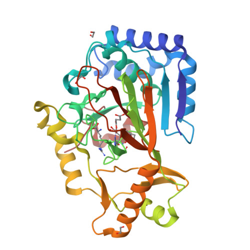
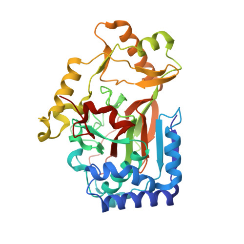
Two Distinct Mechanisms for C-C Desaturation by Iron(II)- and 2-(Oxo)glutarate-Dependent Oxygenases: Importance of alpha-Heteroatom Assistance.

Dunham, N.P.Chang, W.C.Mitchell, A.J.Martinie, R.J.Zhang, B.Bergman, J.A.Rajakovich, L.J.Wang, B.Silakov, A.Krebs, C.Boal, A.K.Bollinger, J.M.

(2018) J Am Chem Soc 140: 7116-7126

  • DOI: https://doi.org/10.1021/jacs.8b01933
  • Primary Citation of Related Structures:  
    6DAW, 6DAX, 6DAZ, 6DB2

  • PubMed Abstract: 

    Hydroxylation of aliphatic carbons by nonheme Fe(IV)-oxo (ferryl) complexes proceeds by hydrogen-atom (H•) transfer (HAT) to the ferryl and subsequent coupling between the carbon radical and Fe(III)-coordinated oxygen (termed rebound). Enzymes that use H•-abstracting ferryl complexes for other transformations must either suppress rebound or further process hydroxylated intermediates. For olefin-installing C-C desaturations, it has been proposed that a second HAT to the Fe(III)-OH complex from the carbon α to the radical preempts rebound. Deuterium ( 2 H) at the second site should slow this step, potentially making rebound competitive. Desaturations mediated by two related l-arginine-modifying iron(II)- and 2-(oxo)glutarate-dependent (Fe/2OG) oxygenases behave oppositely in this key test, implicating different mechanisms. NapI, the l-Arg 4,5-desaturase from the naphthyridinomycin biosynthetic pathway, abstracts H• first from C5 but hydroxylates this site (leading to guanidine release) to the same modest extent whether C4 harbors 1 H or 2 H. By contrast, an unexpected 3,4-desaturation of l-homoarginine (l-hArg) by VioC, the l-Arg 3-hydroxylase from the viomycin biosynthetic pathway, is markedly disfavored relative to C4 hydroxylation when C3 (the second hydrogen donor) harbors 2 H. Anchimeric assistance by N6 permits removal of the C4-H as a proton in the NapI reaction, but, with no such assistance possible in the VioC desaturation, a second HAT step (from C3) is required. The close proximity (≤3.5 Å) of both l-hArg carbons to the oxygen ligand in an X-ray crystal structure of VioC harboring a vanadium-based ferryl mimic supports and rationalizes the sequential-HAT mechanism. The results suggest that, although the sequential-HAT mechanism is feasible, its geometric requirements may make competing hydroxylation unavoidable, thus explaining the presence of α-heteroatoms in nearly all native substrates for Fe/2OG desaturases.


Macromolecules
Find similar proteins by:  (by identity cutoff)  |  3D Structure
Entity ID: 1
MoleculeChains Sequence LengthOrganismDetailsImage
Alpha-ketoglutarate-dependent L-arginine hydroxylase336Streptomyces vinaceusMutation(s): 0 
EC: 1.14.11.41
UniProt
Find proteins for Q6WZB0 (Streptomyces vinaceus)
Explore Q6WZB0 
Go to UniProtKB:  Q6WZB0
Entity Groups  
Sequence Clusters30% Identity50% Identity70% Identity90% Identity95% Identity100% Identity
UniProt GroupQ6WZB0
Sequence Annotations
Expand
  • Reference Sequence
Small Molecules
Ligands 4 Unique
IDChains Name / Formula / InChI Key2D Diagram3D Interactions
HRG
Query on HRG

Download Ideal Coordinates CCD File 
C [auth A]L-HOMOARGININE
C7 H16 N4 O2
QUOGESRFPZDMMT-YFKPBYRVSA-N
SIN
Query on SIN

Download Ideal Coordinates CCD File 
B [auth A]SUCCINIC ACID
C4 H6 O4
KDYFGRWQOYBRFD-UHFFFAOYSA-N
VVO
Query on VVO

Download Ideal Coordinates CCD File 
D [auth A]oxovanadium(2+)
O V
MHHDXUNFNAZUGB-UHFFFAOYSA-N
EDO
Query on EDO

Download Ideal Coordinates CCD File 
E [auth A],
F [auth A],
G [auth A]
1,2-ETHANEDIOL
C2 H6 O2
LYCAIKOWRPUZTN-UHFFFAOYSA-N
Experimental Data & Validation

Experimental Data

  • Method: X-RAY DIFFRACTION
  • Resolution: 1.70 Å
  • R-Value Free:  0.200 (Depositor), 0.210 (DCC) 
  • R-Value Work:  0.175 (Depositor), 0.190 (DCC) 
  • R-Value Observed: 0.176 (Depositor) 
Space Group: C 1 2 1
Unit Cell:
Length ( Å )Angle ( ˚ )
a = 81.025α = 90
b = 66.637β = 109.47
c = 63.224γ = 90
Software Package:
Software NamePurpose
REFMACrefinement
HKL-2000data scaling
PDB_EXTRACTdata extraction
HKL-2000data reduction
PHASERphasing

Structure Validation

View Full Validation Report


Ligand Structure Quality Assessment 

Created with Raphaël 2.3.0Worse 01 BetterLigand structure goodness of fit to experimental dataBest fitted HRGClick on this verticalbar to view details

Entry History & Funding Information

Deposition Data


Funding OrganizationLocationGrant Number
National Institutes of Health/National Institute of General Medical Sciences (NIH/NIGMS)United StatesGM119707

Revision History  (Full details and data files)

  • Version 1.0: 2018-05-16
    Type: Initial release
  • Version 1.1: 2018-06-20
    Changes: Data collection, Database references
  • Version 1.2: 2020-01-01
    Changes: Author supporting evidence
  • Version 1.3: 2024-03-13
    Changes: Data collection, Database references