6FKW

Europium-containing methanol dehydrogenase


Experimental Data Snapshot

  • Method: X-RAY DIFFRACTION
  • Resolution: 1.40 Å
  • R-Value Free: 0.185 
  • R-Value Work: 0.169 
  • R-Value Observed: 0.169 

wwPDB Validation   3D Report Full Report


Ligand Structure Quality Assessment 


This is version 1.1 of the entry. See complete history


Literature

Similar but not the same: First Kinetic and Structural Analyses of a Methanol Dehydrogenase Containing a Europium Ion in the Active Site.

Jahn, B.Pol, A.Lumpe, H.Barends, T.Dietl, A.Hogendoorn, C.Op den Camp, H.Daumann, L.

(2018) Chembiochem 

  • DOI: https://doi.org/10.1002/cbic.201800130
  • Primary Citation of Related Structures:  
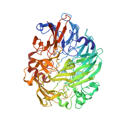
    6FKW

  • PubMed Abstract: 

    Since the discovery of the biological relevance of rare earth elements (REEs) for numerous different bacteria, questions concerning the advantages of REEs in the active sites of methanol dehydrogenases (MDHs) over calcium(II) and of why bacteria prefer light REEs have been a subject of debate. Here we report the cultivation and purification of the strictly REE-dependent methanotrophic bacterium Methylacidiphilum fumariolicum SolV with europium(III), as well as structural and kinetic analyses of the first methanol dehydrogenase incorporating Eu in the active site. Crystal structure determination of the Eu-MDH demonstrated that overall no major structural changes were induced by conversion to this REE. Circular dichroism (CD) measurements were used to determine optimal conditions for kinetic assays, whereas inductively coupled plasma mass spectrometry (ICP-MS) showed 70 % incorporation of Eu in the enzyme. Our studies explain why bacterial growth of SolV in the presence of Eu 3+ is significantly slower than in the presence of La 3+ /Ce 3+ /Pr 3+ : Eu-MDH possesses a decreased catalytic efficiency. Although REEs have similar properties, the differences in ionic radii and coordination numbers across the series significantly impact MDH efficiency.


  • Organizational Affiliation

    Ludwig-Maximilians-Universität München, Department Chemie, Butenandtstr. 5-13, 81377, München, Germany.


Macromolecules
Find similar proteins by:  (by identity cutoff)  |  3D Structure
Entity ID: 1
MoleculeChains Sequence LengthOrganismDetailsImage
Methanol dehydrogenase
A, B, C, D
577Methylacidiphilum fumariolicum SolVMutation(s): 0 
EC: 1.1.2.7 (PDB Primary Data), 1.1.2.8 (PDB Primary Data)
UniProt
Find proteins for I0JWN7 (Methylacidiphilum fumariolicum (strain SolV))
Explore I0JWN7 
Go to UniProtKB:  I0JWN7
Entity Groups  
Sequence Clusters30% Identity50% Identity70% Identity90% Identity95% Identity100% Identity
UniProt GroupI0JWN7
Sequence Annotations
Expand
  • Reference Sequence
Experimental Data & Validation

Experimental Data

  • Method: X-RAY DIFFRACTION
  • Resolution: 1.40 Å
  • R-Value Free: 0.185 
  • R-Value Work: 0.169 
  • R-Value Observed: 0.169 
  • Space Group: P 1
Unit Cell:
Length ( Å )Angle ( ˚ )
a = 55.08α = 115.21
b = 101.18β = 97.53
c = 107.79γ = 91.78
Software Package:
Software NamePurpose
REFMACrefinement
XDSdata reduction
XSCALEdata scaling
REFMACphasing

Structure Validation

View Full Validation Report


Ligand Structure Quality Assessment 


Entry History & Funding Information

Deposition Data


Funding OrganizationLocationGrant Number
ERCNetherlandsERC Advanced Grant project VOLCANO 669371

Revision History  (Full details and data files)

  • Version 1.0: 2018-03-21
    Type: Initial release
  • Version 1.1: 2024-01-17
    Changes: Data collection, Database references, Derived calculations, Refinement description