6HKQ | pdb_00006hkq

Human GPX4 in complex with covalent Inhibitor ML162 (S enantiomer)


Experimental Data Snapshot

  • Method: X-RAY DIFFRACTION
  • Resolution: 1.54 Å
  • R-Value Free: 
    0.184 (Depositor), 0.190 (DCC) 
  • R-Value Work: 
    0.149 (Depositor), 0.150 (DCC) 
  • R-Value Observed: 
    0.151 (Depositor) 

wwPDB Validation   3D Report Full Report


Ligand Structure Quality Assessment 

Created with Raphaël 2.3.0Worse 01 BetterLigand structure goodness of fit to experimental dataBest fitted G9NClick on this verticalbar to view details

This is version 1.2 of the entry. See complete history


Literature

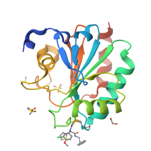
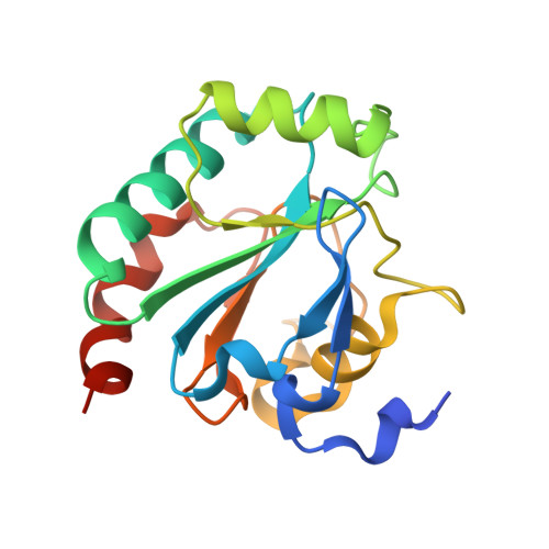
Crystal structures of the selenoprotein glutathione peroxidase 4 in its apo form and in complex with the covalently bound inhibitor ML162.

Moosmayer, D.Hilpmann, A.Hoffmann, J.Schnirch, L.Zimmermann, K.Badock, V.Furst, L.Eaton, J.K.Viswanathan, V.S.Schreiber, S.L.Gradl, S.Hillig, R.C.

(2021) Acta Crystallogr D Struct Biol 77: 237-248

  • DOI: https://doi.org/10.1107/S2059798320016125
  • Primary Citation of Related Structures:  
    6HKQ, 6HN3

  • PubMed Abstract: 

    Wild-type human glutathione peroxidase 4 (GPX4) was co-expressed with SBP2 (selenocysteine insertion sequence-binding protein 2) in human HEK cells to achieve efficient production of this selenocysteine-containing enzyme on a preparative scale for structural biology. The protein was purified and crystallized, and the crystal structure of the wild-type form of GPX4 was determined at 1.0 Å resolution. The overall fold and the active site are conserved compared with previously determined crystal structures of mutated forms of GPX4. A mass-spectrometry-based approach was developed to monitor the reaction of the active-site selenocysteine Sec46 with covalent inhibitors. This, together with the introduction of a surface mutant (Cys66Ser), enabled the crystal structure determination of GPX4 in complex with the covalent inhibitor ML162 [(S)-enantiomer]. The mass-spectrometry-based approach described here opens the path to further co-complex crystal structures of this potential cancer drug target in complex with covalent inhibitors.


  • Organizational Affiliation

    Research and Development, Pharmaceuticals, Bayer AG, 13353 Berlin, Germany.


Macromolecules
Find similar proteins by:  (by identity cutoff)  |  3D Structure
Entity ID: 1
MoleculeChains Sequence LengthOrganismDetailsImage
Phospholipid hydroperoxide glutathione peroxidase176Homo sapiensMutation(s): 1 
Gene Names: GPX4
EC: 1.11.1.12 (PDB Primary Data), 1.11.1.9 (UniProt)
Membrane Entity: Yes 
UniProt & NIH Common Fund Data Resources
Find proteins for P36969 (Homo sapiens)
Explore P36969 
Go to UniProtKB:  P36969
PHAROS:  P36969
GTEx:  ENSG00000167468 
Entity Groups  
Sequence Clusters30% Identity50% Identity70% Identity90% Identity95% Identity100% Identity
UniProt GroupP36969
Sequence Annotations
Expand
  • Reference Sequence
Small Molecules
Ligands 4 Unique
IDChains Name / Formula / InChI Key2D Diagram3D Interactions
G9N (Subject of Investigation/LOI)
Query on G9N

Download Ideal Coordinates CCD File 
B [auth A](2~{S})-2-[2-chloranylethanoyl-(3-chloranyl-4-methoxy-phenyl)amino]-~{N}-(2-phenylethyl)-2-thiophen-2-yl-ethanamide
C23 H22 Cl2 N2 O3 S
UNVKYJSNMVDZJE-JOCHJYFZSA-N
SO4
Query on SO4

Download Ideal Coordinates CCD File 
C [auth A]SULFATE ION
O4 S
QAOWNCQODCNURD-UHFFFAOYSA-L
DMS
Query on DMS

Download Ideal Coordinates CCD File 
E [auth A],
F [auth A]
DIMETHYL SULFOXIDE
C2 H6 O S
IAZDPXIOMUYVGZ-UHFFFAOYSA-N
EDO
Query on EDO

Download Ideal Coordinates CCD File 
D [auth A]1,2-ETHANEDIOL
C2 H6 O2
LYCAIKOWRPUZTN-UHFFFAOYSA-N
Binding Affinity Annotations 
IDSourceBinding Affinity
G9N BindingDB:  6HKQ Ki: 2.89e+4 (nM) from 1 assay(s)
IC50: 4850 (nM) from 1 assay(s)
EC50: min: 28, max: 2200 (nM) from 2 assay(s)
Experimental Data & Validation

Experimental Data

  • Method: X-RAY DIFFRACTION
  • Resolution: 1.54 Å
  • R-Value Free:  0.184 (Depositor), 0.190 (DCC) 
  • R-Value Work:  0.149 (Depositor), 0.150 (DCC) 
  • R-Value Observed: 0.151 (Depositor) 
Space Group: P 21 21 21
Unit Cell:
Length ( Å )Angle ( ˚ )
a = 32.734α = 90
b = 57.249β = 90
c = 81.311γ = 90
Software Package:
Software NamePurpose
REFMACrefinement
HKL-3000data reduction
HKL-3000data scaling
PHASERphasing

Structure Validation

View Full Validation Report


Ligand Structure Quality Assessment 

Created with Raphaël 2.3.0Worse 01 BetterLigand structure goodness of fit to experimental dataBest fitted G9NClick on this verticalbar to view details

Entry History 

Deposition Data

Revision History  (Full details and data files)

  • Version 1.0: 2020-04-01
    Type: Initial release
  • Version 1.1: 2021-06-16
    Changes: Database references
  • Version 1.2: 2022-12-07
    Changes: Advisory, Database references, Derived calculations, Source and taxonomy