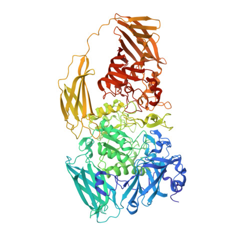
6KUZ

E.coli beta-galactosidase (E537Q) in complex with fluorescent probe KSL01


Experimental Data Snapshot

  • Method: X-RAY DIFFRACTION
  • Resolution: 2.83 Å
  • R-Value Free: 0.224 
  • R-Value Work: 0.182 
  • R-Value Observed: 0.184 

wwPDB Validation   3D Report Full Report


Ligand Structure Quality Assessment 


This is version 1.2 of the entry. See complete history


Literature

First-generation species-selective chemical probes for fluorescence imaging of human senescence-associated beta-galactosidase.

Li, X.Qiu, W.Li, J.Chen, X.Hu, Y.Gao, Y.Shi, D.Li, X.Lin, H.Hu, Z.Dong, G.Sheng, C.Jiang, B.Xia, C.Kim, C.Y.Guo, Y.Li, J.

(2020) Chem Sci 11: 7292-7301

  • DOI: https://doi.org/10.1039/d0sc01234c
  • Primary Citation of Related Structures:  
    6KUZ

  • PubMed Abstract: 

    Human senescence-associated β-galactosidase (SA-β-gal), the most widely used biomarker of aging, is a valuable tool for assessing the extent of cell 'healthy aging' and potentially predicting the health life span of an individual. Human SA-β-gal is an endogenous lysosomal enzyme expressed from GLB1 , the catalytic domain of which is very different from that of E. coli β-gal, a bacterial enzyme encoded by lacZ . However, existing chemical probes for this marker still lack the ability to distinguish human SA-β-gal from β-gal of other species, such as bacterial β-gal, which can yield false positive signals. Here, we show a molecular design strategy to construct fluorescent probes with the above ability with the aid of structure-based steric hindrance adjustment catering to different enzyme pockets. The resulting probes normally work as traditional SA-β-gal probes, but they are unique in their powerful ability to distinguish human SA-β-gal from E. coli β-gal, thus achieving species-selective visualization of human SA-β-gal for the first time. NIR-emitting fluorescent probe KSL11 as their representative further displays excellent species-selective recognition performance in biological systems, which has been herein verified by testing in senescent cells, in lacZ -transfected cells and in E. coli -β-gal-contaminated tissue sections of mice. Because of our probes, it was also discovered that SA-β-gal content in mice increased gradually with age and SA-β-gal accumulated most in the kidneys among the main organs of naturally aging mice, suggesting that the kidneys are the organs with the most severe aging during natural aging.


  • Organizational Affiliation

    State Key Laboratory of Bioreactor Engineering, Shanghai Key Laboratory of New Drug Design, School of Pharmacy, East China University of Science and Technology Shanghai 200237 China jianli@ecust.edu.cn.


Macromolecules
Find similar proteins by:  (by identity cutoff)  |  3D Structure
Entity ID: 1
MoleculeChains Sequence LengthOrganismDetailsImage
Beta-galactosidase
A, B, C, D
1,024Escherichia coli K-12Mutation(s): 1 
Gene Names: lacZb0344JW0335
EC: 3.2.1.23
UniProt
Find proteins for P00722 (Escherichia coli (strain K12))
Explore P00722 
Go to UniProtKB:  P00722
Entity Groups  
Sequence Clusters30% Identity50% Identity70% Identity90% Identity95% Identity100% Identity
UniProt GroupP00722
Sequence Annotations
Expand
  • Reference Sequence
Small Molecules
Ligands 5 Unique
IDChains Name / Formula / InChI Key2D Diagram3D Interactions
DVL (Subject of Investigation/LOI)
Query on DVL

Download Ideal Coordinates CCD File 
E [auth A],
LA [auth C],
WA [auth D],
X [auth B]
3-(1,3-benzothiazol-2-yl)-2-[[4-[(2~{S},3~{R},4~{S},5~{R},6~{R})-6-(hydroxymethyl)-3,4,5-tris(oxidanyl)oxan-2-yl]oxyphenyl]methoxy]-5-methyl-benzaldehyde
C28 H27 N O8 S
OIAKZSZIDGSPEQ-WRPMCVFTSA-N
GOL
Query on GOL

Download Ideal Coordinates CCD File 
JB [auth D]GLYCEROL
C3 H8 O3
PEDCQBHIVMGVHV-UHFFFAOYSA-N
DMS
Query on DMS

Download Ideal Coordinates CCD File 
AB [auth D]
BA [auth B]
BB [auth D]
CA [auth B]
CB [auth D]
AB [auth D],
BA [auth B],
BB [auth D],
CA [auth B],
CB [auth D],
DA [auth B],
DB [auth D],
EA [auth B],
EB [auth D],
FA [auth B],
FB [auth D],
GA [auth B],
GB [auth D],
HA [auth B],
HB [auth D],
I [auth A],
IA [auth B],
IB [auth D],
J [auth A],
K [auth A],
L [auth A],
M [auth A],
N [auth A],
O [auth A],
P [auth A],
PA [auth C],
Q [auth A],
QA [auth C],
R [auth A],
RA [auth C],
S [auth A],
SA [auth C],
T [auth A],
TA [auth C],
U [auth A]
DIMETHYL SULFOXIDE
C2 H6 O S
IAZDPXIOMUYVGZ-UHFFFAOYSA-N
MG
Query on MG

Download Ideal Coordinates CCD File 
JA [auth B]
KA [auth B]
KB [auth D]
LB [auth D]
UA [auth C]
JA [auth B],
KA [auth B],
KB [auth D],
LB [auth D],
UA [auth C],
V [auth A],
VA [auth C],
W [auth A]
MAGNESIUM ION
Mg
JLVVSXFLKOJNIY-UHFFFAOYSA-N
NA
Query on NA

Download Ideal Coordinates CCD File 
AA [auth B]
F [auth A]
G [auth A]
H [auth A]
MA [auth C]
AA [auth B],
F [auth A],
G [auth A],
H [auth A],
MA [auth C],
NA [auth C],
OA [auth C],
XA [auth D],
Y [auth B],
YA [auth D],
Z [auth B],
ZA [auth D]
SODIUM ION
Na
FKNQFGJONOIPTF-UHFFFAOYSA-N
Experimental Data & Validation

Experimental Data

  • Method: X-RAY DIFFRACTION
  • Resolution: 2.83 Å
  • R-Value Free: 0.224 
  • R-Value Work: 0.182 
  • R-Value Observed: 0.184 
  • Space Group: C 1 2 1
Unit Cell:
Length ( Å )Angle ( ˚ )
a = 248.174α = 90
b = 85.506β = 94.09
c = 243.052γ = 90
Software Package:
Software NamePurpose
REFMACrefinement
HKL-2000data reduction
Aimlessdata scaling
MOLREPphasing

Structure Validation

View Full Validation Report


Ligand Structure Quality Assessment 


Entry History 

Deposition Data

Revision History  (Full details and data files)

  • Version 1.0: 2020-07-08
    Type: Initial release
  • Version 1.1: 2021-06-30
    Changes: Database references, Derived calculations
  • Version 1.2: 2023-11-22
    Changes: Data collection, Database references, Refinement description