6RLX

X-RAY STRUCTURE OF HUMAN RELAXIN AT 1.5 ANGSTROMS. COMPARISON TO INSULIN AND IMPLICATIONS FOR RECEPTOR BINDING DETERMINANTS


Experimental Data Snapshot

  • Method: X-RAY DIFFRACTION
  • Resolution: 1.50 Å
  • R-Value Work: 0.187 
  • R-Value Observed: 0.187 

wwPDB Validation   3D Report Full Report


This is version 2.1 of the entry. See complete history


Literature

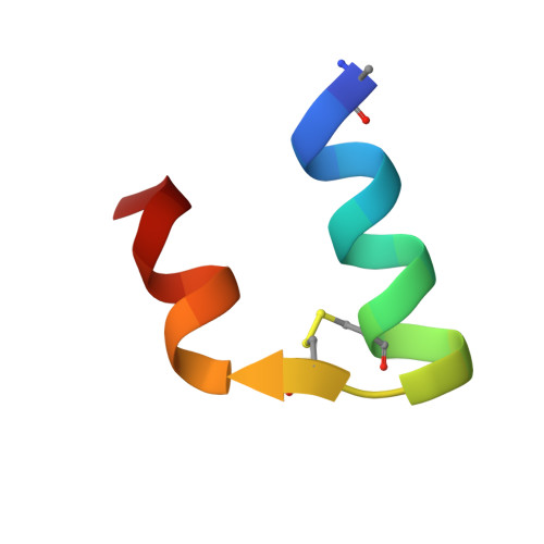
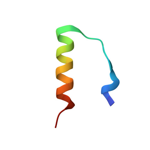
X-ray structure of human relaxin at 1.5 A. Comparison to insulin and implications for receptor binding determinants.

Eigenbrot, C.Randal, M.Quan, C.Burnier, J.O'Connell, L.Rinderknecht, E.Kossiakoff, A.A.

(1991) J Mol Biol 221: 15-21

  • Primary Citation of Related Structures:  
    6RLX

  • PubMed Abstract: 

    The X-ray crystal structure of relaxin at 1.5 A resolution is reported for the physiologically active form of the human hormone. Relaxin is a small, two-chain polypeptide that is a member of the protein hormone family that also includes insulin and the insulin-like growth factors IGF-I and IGF-II. These hormones have biologically diverse activities but are structurally similar, sharing a distinctive pattern of cysteine and glycine residues. The predicted structural homology of relaxin to insulin is confirmed by this structural analysis; however, there are significant differences in the terminal regions of the b-chain. Although relaxin, like insulin, crystallizes as a dimer, the orientation of the molecules in the respective dimers is completely different. The region of the relaxin molecule proposed to be involved in receptor binding is part of the dimer interface, suggesting that some of the other residues contained in the dimer contact surface might be receptor binding determinants as well. The proposed receptor binding determinants for insulin likewise include residues at its dimer interface. However, because the dimer contacts of relaxin and insulin are quite different, it appears that these two structurally related hormones have evolved somewhat dissimilar mechanisms for receptor binding.


  • Organizational Affiliation

    Protein Engineering, Bioorganic Chemistry Department, Genentech, Inc., South San Francisco, CA 94080.


Macromolecules

Find similar proteins by:  Sequence   |   3D Structure  

Entity ID: 1
MoleculeChains Sequence LengthOrganismDetailsImage
RELAXIN, A-CHAIN
A, C
24Homo sapiensMutation(s): 0 
UniProt & NIH Common Fund Data Resources
Find proteins for P04090 (Homo sapiens)
Explore P04090 
Go to UniProtKB:  P04090
PHAROS:  P04090
GTEx:  ENSG00000107014 
Entity Groups  
Sequence Clusters30% Identity50% Identity70% Identity90% Identity95% Identity100% Identity
UniProt GroupP04090
Sequence Annotations
Expand
  • Reference Sequence
Find similar proteins by:  (by identity cutoff)  |  3D Structure
Entity ID: 2
MoleculeChains Sequence LengthOrganismDetailsImage
RELAXIN, B-CHAIN
B, D
28Homo sapiensMutation(s): 0 
UniProt & NIH Common Fund Data Resources
Find proteins for P04090 (Homo sapiens)
Explore P04090 
Go to UniProtKB:  P04090
PHAROS:  P04090
GTEx:  ENSG00000107014 
Entity Groups  
Sequence Clusters30% Identity50% Identity70% Identity90% Identity95% Identity100% Identity
UniProt GroupP04090
Sequence Annotations
Expand
  • Reference Sequence
Small Molecules
Modified Residues  1 Unique
IDChains TypeFormula2D DiagramParent
PCA
Query on PCA
A, C
L-PEPTIDE LINKINGC5 H7 N O3GLN
Experimental Data & Validation

Experimental Data

  • Method: X-RAY DIFFRACTION
  • Resolution: 1.50 Å
  • R-Value Work: 0.187 
  • R-Value Observed: 0.187 
  • Space Group: C 2 2 21
Unit Cell:
Length ( Å )Angle ( ˚ )
a = 53.97α = 90
b = 56.88β = 90
c = 61.68γ = 90
Software Package:
Software NamePurpose
X-PLORmodel building
PROLSQrefinement
X-PLORrefinement
X-PLORphasing

Structure Validation

View Full Validation Report


Entry History 

Deposition Data

Revision History  (Full details and data files)

  • Version 1.0: 1993-10-31
    Type: Initial release
  • Version 1.1: 2008-03-25
    Changes: Version format compliance
  • Version 1.2: 2011-07-13
    Changes: Version format compliance
  • Version 1.3: 2011-11-16
    Changes: Atomic model
  • Version 1.4: 2019-07-17
    Changes: Data collection, Derived calculations, Other, Refinement description
  • Version 1.5: 2019-08-14
    Changes: Data collection, Refinement description
  • Version 2.0: 2019-12-25
    Changes: Derived calculations, Polymer sequence
  • Version 2.1: 2024-03-13
    Changes: Data collection, Database references, Source and taxonomy