6V9Z

Cryo-EM structure of PCAT1 bound to its CtA peptide substrate


Experimental Data Snapshot

  • Method: ELECTRON MICROSCOPY
  • Resolution: 3.35 Å
  • Aggregation State: PARTICLE 
  • Reconstruction Method: SINGLE PARTICLE 

wwPDB Validation   3D Report Full Report


This is version 1.2 of the entry. See complete history


Literature

Structural basis of substrate recognition by a polypeptide processing and secretion transporter.

Kieuvongngam, V.Olinares, P.D.B.Palillo, A.Oldham, M.L.Chait, B.T.Chen, J.

(2020) Elife 9

  • DOI: https://doi.org/10.7554/eLife.51492
  • Primary Citation of Related Structures:  
    6V9Z

  • PubMed Abstract: 

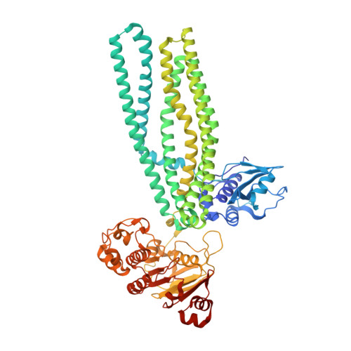
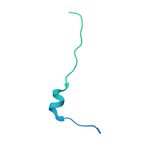
    The peptidase-containing ATP-binding cassette transporters (PCATs) are unique members of the ABC transporter family that proteolytically process and export peptides and proteins. Each PCAT contains two peptidase domains that cleave off the secretion signal, two transmembrane domains forming a translocation pathway, and two nucleotide-binding domains that hydrolyze ATP. Previously the crystal structures of a PCAT from Clostridium thermocellum (PCAT1) were determined in the absence and presence of ATP, revealing how ATP binding regulates the protease activity and access to the translocation pathway. However, how the substrate CtA, a 90-residue polypeptide, is recognized by PCAT1 remained elusive. To address this question, we determined the structure of the PCAT1-CtA complex by electron cryo-microscopy (cryo-EM) to 3.4 Å resolution. The structure shows that two CtAs are bound via their N-terminal leader peptides, but only one is positioned for cleavage and translocation. Based on these results, we propose a model of how substrate cleavage, ATP hydrolysis, and substrate translocation are coordinated in a transport cycle.


  • Organizational Affiliation

    Laboratory of Membrane Biophysics and Biology, The Rockefeller University, New York, United States.


Macromolecules
Find similar proteins by:  (by identity cutoff)  |  3D Structure
Entity ID: 1
MoleculeChains Sequence LengthOrganismDetailsImage
ABC-type bacteriocin transporterA,
C [auth B]
730Acetivibrio thermocellus ATCC 27405Mutation(s): 1 
Gene Names: Cthe_0534
Membrane Entity: Yes 
UniProt
Find proteins for A3DCU1 (Acetivibrio thermocellus (strain ATCC 27405 / DSM 1237 / JCM 9322 / NBRC 103400 / NCIMB 10682 / NRRL B-4536 / VPI 7372))
Explore A3DCU1 
Go to UniProtKB:  A3DCU1
Entity Groups  
Sequence Clusters30% Identity50% Identity70% Identity90% Identity95% Identity100% Identity
UniProt GroupA3DCU1
Sequence Annotations
Expand
  • Reference Sequence
Find similar proteins by:  (by identity cutoff)  |  3D Structure
Entity ID: 2
MoleculeChains Sequence LengthOrganismDetailsImage
CtAB [auth C],
D
93Acetivibrio thermocellus ATCC 27405Mutation(s): 0 
Gene Names: Cthe_0535
UniProt
Find proteins for A3DCU2 (Acetivibrio thermocellus (strain ATCC 27405 / DSM 1237 / JCM 9322 / NBRC 103400 / NCIMB 10682 / NRRL B-4536 / VPI 7372))
Explore A3DCU2 
Go to UniProtKB:  A3DCU2
Entity Groups  
Sequence Clusters30% Identity50% Identity70% Identity90% Identity95% Identity100% Identity
UniProt GroupA3DCU2
Sequence Annotations
Expand
  • Reference Sequence
Experimental Data & Validation

Experimental Data

  • Method: ELECTRON MICROSCOPY
  • Resolution: 3.35 Å
  • Aggregation State: PARTICLE 
  • Reconstruction Method: SINGLE PARTICLE 
EM Software:
TaskSoftware PackageVersion
RECONSTRUCTIONcryoSPARCv.0.6.5
MODEL REFINEMENTCoot0.8.9.2

Structure Validation

View Full Validation Report


Entry History & Funding Information

Deposition Data


Funding OrganizationLocationGrant Number
Howard Hughes Medical Institute (HHMI)United States--

Revision History  (Full details and data files)

  • Version 1.0: 2020-01-22
    Type: Initial release
  • Version 1.1: 2020-01-29
    Changes: Database references
  • Version 1.2: 2024-03-06
    Changes: Data collection, Database references, Refinement description