6Y4Q

Structure of a stapled peptide bound to MDM2


Experimental Data Snapshot

  • Method: X-RAY DIFFRACTION
  • Resolution: 1.63 Å
  • R-Value Free: 0.229 
  • R-Value Work: 0.194 
  • R-Value Observed: 0.196 

wwPDB Validation   3D Report Full Report


Ligand Structure Quality Assessment 


This is version 1.2 of the entry. See complete history


Literature

Diarylethene moiety as an enthalpy-entropy switch: photoisomerizable stapled peptides for modulating p53/MDM2 interaction.

Strizhak, A.V.Babii, O.Afonin, S.Bakanovich, I.Pantelejevs, T.Xu, W.Fowler, E.Eapen, R.Sharma, K.Platonov, M.O.Hurmach, V.V.Itzhaki, L.Hyvonen, M.Ulrich, A.S.Spring, D.R.Komarov, I.V.

(2020) Org Biomol Chem 18: 5359-5369

  • DOI: https://doi.org/10.1039/d0ob00831a
  • Primary Citation of Related Structures:  
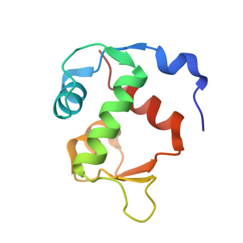
    6Y4Q

  • PubMed Abstract: 

    Analogs of the known inhibitor (peptide pDI) of the p53/MDM2 protein-protein interaction are reported, which are stapled by linkers bearing a photoisomerizable diarylethene moiety. The corresponding photoisomers possess significantly different affinities to the p53-interacting domain of the human MDM2. Apparent dissociation constants are in the picomolar-to-low nanomolar range for those isomers with diarylethene in the "open" configuration, but up to eight times larger for the corresponding "closed" isomers. Spectroscopic, structural, and computational studies showed that the stapling linkers of the peptides contribute to their binding. Calorimetry revealed that the binding of the "closed" isomers is mostly enthalpy-driven, whereas the "open" photoforms bind to the protein stronger due to their increased binding entropy. The results suggest that conformational dynamics of the protein-peptide complexes may explain the differences in the thermodynamic profiles of the binding.


  • Organizational Affiliation

    University Chemical Laboratory, University of Cambridge, Lensfield Road, CB2 1EW Cambridge, UK. Spring@ch.cam.ac.uk and Enamine Ltd, Vul. Chervonotkatska 78, 02094 Kyiv, Ukraine.


Macromolecules
Find similar proteins by:  (by identity cutoff)  |  3D Structure
Entity ID: 1
MoleculeChains Sequence LengthOrganismDetailsImage
E3 ubiquitin-protein ligase Mdm2
A, B
92Homo sapiensMutation(s): 2 
Gene Names: MDM2
EC: 2.3.2.27
UniProt & NIH Common Fund Data Resources
Find proteins for Q00987 (Homo sapiens)
Explore Q00987 
Go to UniProtKB:  Q00987
PHAROS:  Q00987
GTEx:  ENSG00000135679 
Entity Groups  
Sequence Clusters30% Identity50% Identity70% Identity90% Identity95% Identity100% Identity
UniProt GroupQ00987
Sequence Annotations
Expand
  • Reference Sequence

Find similar proteins by:  Sequence   |   3D Structure  

Entity ID: 2
MoleculeChains Sequence LengthOrganismDetailsImage
ACE-LEU-THR-PHE-GLY-GLU-TYR-TRP-ALA-GLN-LEU-ALA-SER
C, D
13Homo sapiensMutation(s): 0 
Entity Groups  
Sequence Clusters30% Identity50% Identity70% Identity90% Identity95% Identity100% Identity
Sequence Annotations
Expand
  • Reference Sequence
Small Molecules
Ligands 1 Unique
IDChains Name / Formula / InChI Key2D Diagram3D Interactions
O9E (Subject of Investigation/LOI)
Query on O9E

Download Ideal Coordinates CCD File 
E [auth C],
F [auth D]
~{N}-[(1-ethyl-1,2,3-triazol-4-yl)methyl]-~{N},5-dimethyl-4-[2-[2-methyl-5-[methyl-[(1-propyl-1,2,3-triazol-4-yl)methyl]carbamoyl]thiophen-3-yl]cyclopenten-1-yl]thiophene-2-carboxamide
C30 H38 N8 O2 S2
GGDTYYUIMGUGDO-UHFFFAOYSA-N
Experimental Data & Validation

Experimental Data

  • Method: X-RAY DIFFRACTION
  • Resolution: 1.63 Å
  • R-Value Free: 0.229 
  • R-Value Work: 0.194 
  • R-Value Observed: 0.196 
  • Space Group: P 1
Unit Cell:
Length ( Å )Angle ( ˚ )
a = 34.24α = 93.38
b = 34.84β = 107.38
c = 48.41γ = 117.45
Software Package:
Software NamePurpose
Aimlessdata scaling
BUSTERrefinement
PDB_EXTRACTdata extraction
XDSdata reduction
PHASERphasing

Structure Validation

View Full Validation Report


Ligand Structure Quality Assessment 


Entry History & Funding Information

Deposition Data


Funding OrganizationLocationGrant Number
Medical Research Council (MRC, United Kingdom)United Kingdom--

Revision History  (Full details and data files)

  • Version 1.0: 2020-05-20
    Type: Initial release
  • Version 1.1: 2020-07-29
    Changes: Database references
  • Version 1.2: 2024-01-24
    Changes: Advisory, Data collection, Database references, Refinement description