6AY2

Structure of CathB with covalently linked Compound 28


Experimental Data Snapshot

  • Method: X-RAY DIFFRACTION
  • Resolution: 1.60 Å
  • R-Value Free: 0.176 
  • R-Value Work: 0.161 
  • R-Value Observed: 0.162 

wwPDB Validation   3D Report Full Report


Ligand Structure Quality Assessment 


This is version 1.1 of the entry. See complete history


Literature

Discovery of Peptidomimetic Antibody-Drug Conjugate Linkers with Enhanced Protease Specificity.

Wei, B.Gunzner-Toste, J.Yao, H.Wang, T.Wang, J.Xu, Z.Chen, J.Wai, J.Nonomiya, J.Tsai, S.P.Chuh, J.Kozak, K.R.Liu, Y.Yu, S.F.Lau, J.Li, G.Phillips, G.D.Leipold, D.Kamath, A.Su, D.Xu, K.Eigenbrot, C.Steinbacher, S.Ohri, R.Raab, H.Staben, L.R.Zhao, G.Flygare, J.A.Pillow, T.H.Verma, V.Masterson, L.A.Howard, P.W.Safina, B.

(2018) J Med Chem 61: 989-1000

  • DOI: https://doi.org/10.1021/acs.jmedchem.7b01430
  • Primary Citation of Related Structures:  
    6AY2

  • PubMed Abstract: 

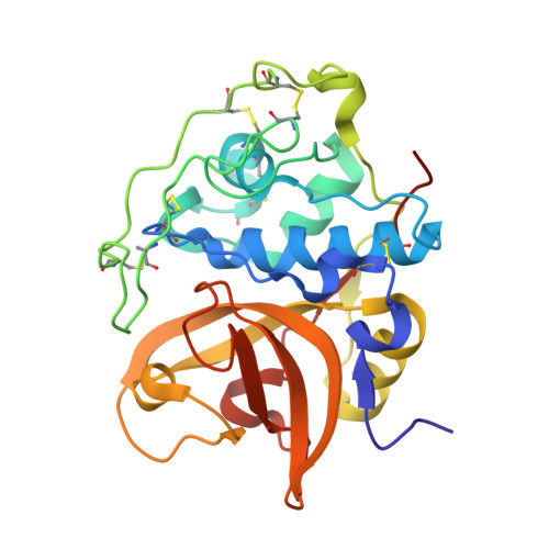
    Antibody-drug conjugates (ADCs) have become an important therapeutic modality for oncology, with three approved by the FDA and over 60 others in clinical trials. Despite the progress, improvements in ADC therapeutic index are desired. Peptide-based ADC linkers that are cleaved by lysosomal proteases have shown sufficient stability in serum and effective payload-release in targeted cells. If the linker can be preferentially hydrolyzed by tumor-specific proteases, safety margin may improve. However, the use of peptide-based linkers limits our ability to modulate protease specificity. Here we report the structure-guided discovery of novel, nonpeptidic ADC linkers. We show that a cyclobutane-1,1-dicarboxamide-containing linker is hydrolyzed predominantly by cathepsin B while the valine-citrulline dipeptide linker is not. ADCs bearing the nonpeptidic linker are as efficacious and stable in vivo as those with the dipeptide linker. Our results strongly support the application of the peptidomimetic linker and present new opportunities for improving the selectivity of ADCs.


  • Organizational Affiliation

    Genentech, Inc. , 1 DNA Way, South San Francisco, California 94080, United States.


Macromolecules
Find similar proteins by:  (by identity cutoff)  |  3D Structure
Entity ID: 1
MoleculeChains Sequence LengthOrganismDetailsImage
Cathepsin B
A, B
255Homo sapiensMutation(s): 0 
Gene Names: CTSBCPSB
EC: 3.4.22.1
UniProt & NIH Common Fund Data Resources
Find proteins for P07858 (Homo sapiens)
Explore P07858 
Go to UniProtKB:  P07858
PHAROS:  P07858
GTEx:  ENSG00000164733 
Entity Groups  
Sequence Clusters30% Identity50% Identity70% Identity90% Identity95% Identity100% Identity
UniProt GroupP07858
Sequence Annotations
Expand
  • Reference Sequence
Small Molecules
Ligands 1 Unique
IDChains Name / Formula / InChI Key2D Diagram3D Interactions
C1G (Subject of Investigation/LOI)
Query on C1G

Download Ideal Coordinates CCD File 
C [auth A],
D [auth B]
N~1~-[(2S)-1-amino-5-(carbamoylamino)pentan-2-yl]-N'~1~-[(1R)-1-(thiophen-3-yl)ethyl]cyclobutane-1,1-dicarboxamide
C18 H29 N5 O3 S
QWGHBARYVOPELP-OCCSQVGLSA-N
Experimental Data & Validation

Experimental Data

  • Method: X-RAY DIFFRACTION
  • Resolution: 1.60 Å
  • R-Value Free: 0.176 
  • R-Value Work: 0.161 
  • R-Value Observed: 0.162 
  • Space Group: P 1 21 1
Unit Cell:
Length ( Å )Angle ( ˚ )
a = 31.317α = 90
b = 127.243β = 102.9
c = 70.034γ = 90
Software Package:
Software NamePurpose
XSCALEdata scaling
REFMACrefinement
PDB_EXTRACTdata extraction
XDSdata reduction
PHASERphasing

Structure Validation

View Full Validation Report


Ligand Structure Quality Assessment 


Entry History 

Deposition Data

Revision History  (Full details and data files)

  • Version 1.0: 2017-12-27
    Type: Initial release
  • Version 1.1: 2018-02-21
    Changes: Database references