Reactivity of a fluorine-containing dirhodium tetracarboxylate compound with proteins.
Loreto, D., Esposito, A., Demitri, N., Guaragna, A., Merlino, A.(2022) Dalton Trans 51: 3695-3705
- PubMed: 35166290
- DOI: https://doi.org/10.1039/d2dt00082b
- Primary Citation of Related Structures:
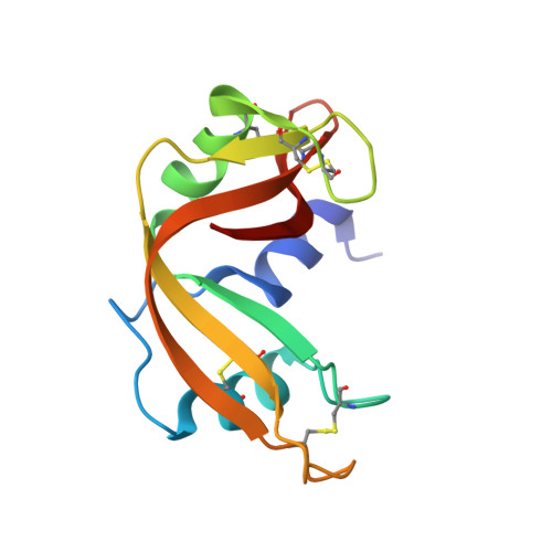
7QPW, 7QPY, 7QPZ, 7QQ0, 7QQ1 - PubMed Abstract:
Dirhodium complexes of general formula [Rh 2 (O 2 CR) 4 ]L 2 are a well-known class of bimetallic compounds that are used as efficient catalysts for a variety of reactions and have been shown to be potent antibacterial and anticancer agents. The catalytic and biological properties of these complexes largely depend on the nature of the bridging carboxylate ligands. Trifluoroacetate (tfa)-containing dirhodium compounds have been used to build artificial metalloenzymes upon reaction with peptides and have been shown to be more cytotoxic than dirhodium tetraacetate. However, there is no structural information on the interaction between these compounds and proteins. Here, cis -Rh 2 (μ-O 2 CCH 3 ) 2 (μ-O 2 CCF 3 ) 2 ([ cis -Rh 2 (OAc) 2 (tfa) 2 ]) has been synthesized and its reaction with bovine pancreatic ribonuclease (RNase A) and hen egg white lysozyme (HEWL) was analyzed using a combination of different techniques, including Fluorine-19 nuclear magnetic resonance spectroscopy and macromolecular X-ray crystallography, with the aim to unveil the differences in the reactivity of tfa-containing dihrodium complexes with proteins when compared to [Rh 2 (OAc) 4 ]. [ cis -Rh 2 (OAc) 2 (tfa) 2 ] and [Rh 2 (OAc) 4 ] bind the N atoms of His side chains of RNase A at the axial position; however the fluorine-containing compound rapidly loses its tfa ligands, while [Rh 2 (OAc) 4 ] can retain the acetate ligands upon protein binding. The reactivity of [ cis -Rh 2 (OAc) 2 (tfa) 2 ] with HEWL is slightly distinct when compared to that of [Rh 2 (OAc) 4 ] under the same experimental conditions; however, both [ cis -Rh 2 (OAc) 2 (tfa) 2 ] and [Rh 2 (OAc) 4 ] degrade when soaked within HEWL crystals. These results provide a structural-based guide for the design of new heterogenous chiral dirhodium/peptide and dirhodium/protein adducts with application in the fields of organic synthesis and asymmetric catalysis.
Organizational Affiliation:
Department of Chemical Sciences, University of Naples Federico II, Complesso Universitario di Monte Sant'Angelo, via Cinthia 21, 80126 Naples, Italy. antonello.merlino@unina.it.