7WAN | pdb_00007wan

Crystal structure of HaloTag complexed with UL2


Experimental Data Snapshot

  • Method: X-RAY DIFFRACTION
  • Resolution: 2.28 Å
  • R-Value Free: 
    0.228 (Depositor), 0.230 (DCC) 
  • R-Value Work: 
    0.178 (Depositor), 0.180 (DCC) 
  • R-Value Observed: 
    0.180 (Depositor) 

Starting Model: experimental
View more details

wwPDB Validation   3D Report Full Report


Ligand Structure Quality Assessment 

Created with Raphaël 2.3.0Worse 01 BetterLigand structure goodness of fit to experimental dataBest fitted 8MSClick on this verticalbar to view details

This is version 1.2 of the entry. See complete history


Literature

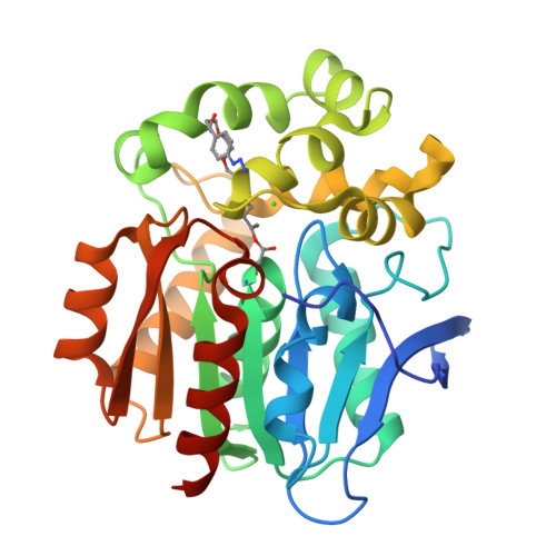
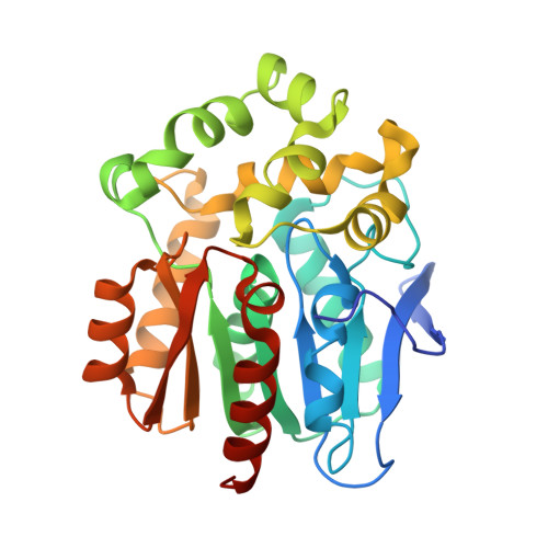
A chemical tool for blue light-inducible proximity photo-crosslinking in live cells.

Mishra, P.K.Kang, M.G.Lee, H.Kim, S.Choi, S.Sharma, N.Park, C.M.Ko, J.Lee, C.Seo, J.K.Rhee, H.W.

(2022) Chem Sci 13: 955-966

  • DOI: https://doi.org/10.1039/d1sc04871f
  • Primary Citation of Related Structures:  
    7WAM, 7WAN

  • PubMed Abstract: 

    We developed a proximity photo-crosslinking method ( Spotlight ) with a 4-azido- N -ethyl-1,8-naphthalimide (AzNP) moiety that can be converted to reactive aryl nitrene species using ambient blue light-emitting diode light. Using an AzNP-conjugated HaloTag ligand (VL1), blue light-induced photo-crosslinked products of various HaloTag-conjugated proteins of interest were detected in subcellular spaces in live cells. Chemical or heat stress-induced dynamic changes in the proteome were also detected, and photo-crosslinking in the mouse brain tissue was enabled. Using Spotlight , we further identified the host interactome of SARS-CoV-2 nucleocapsid (N) protein, which is essential for viral genome assembly. Mass analysis of the VL1-crosslinked product of N -HaloTag in HEK293T cells showed that RNA-binding proteins in stress granules were exclusively enriched in the cross-linked samples. These results tell that our method can reveal the interactome of protein of interest within a short distance in live cells.


  • Organizational Affiliation

    Department of Chemistry, Seoul National University Seoul 08826 Korea rheehw@snu.ac.kr.


Macromolecules
Find similar proteins by:  (by identity cutoff)  |  3D Structure
Entity ID: 1
MoleculeChains Sequence LengthOrganismDetailsImage
Haloalkane dehalogenase299Rhodococcus sp. (in: high G+C Gram-positive bacteria)Mutation(s): 22 
Gene Names: dhaA
EC: 3.8.1.5
UniProt
Find proteins for P0A3G3 (Rhodococcus sp)
Explore P0A3G3 
Go to UniProtKB:  P0A3G3
Entity Groups  
Sequence Clusters30% Identity50% Identity70% Identity90% Identity95% Identity100% Identity
UniProt GroupP0A3G3
Sequence Annotations
Expand
  • Reference Sequence
Small Molecules
Ligands 2 Unique
IDChains Name / Formula / InChI Key2D Diagram3D Interactions
8MS
Query on 8MS

Download Ideal Coordinates CCD File 
B [auth A](R)-[4-(2-azanylhydrazinyl)phenyl]-[2-[2-(2-hexoxyethoxy)ethoxy]ethylamino]methanol
C19 H36 N4 O4
LFWZOPMQWFRAFE-LJQANCHMSA-N
CL
Query on CL

Download Ideal Coordinates CCD File 
C [auth A]CHLORIDE ION
Cl
VEXZGXHMUGYJMC-UHFFFAOYSA-M
Experimental Data & Validation

Experimental Data

  • Method: X-RAY DIFFRACTION
  • Resolution: 2.28 Å
  • R-Value Free:  0.228 (Depositor), 0.230 (DCC) 
  • R-Value Work:  0.178 (Depositor), 0.180 (DCC) 
  • R-Value Observed: 0.180 (Depositor) 
Space Group: P 1 21 1
Unit Cell:
Length ( Å )Angle ( ˚ )
a = 43.906α = 90
b = 72.764β = 109.3
c = 44.432γ = 90
Software Package:
Software NamePurpose
PHENIXrefinement
PDB_EXTRACTdata extraction
HKL-2000data reduction
HKL-2000data scaling
PHENIXphasing

Structure Validation

View Full Validation Report


Ligand Structure Quality Assessment 

Created with Raphaël 2.3.0Worse 01 BetterLigand structure goodness of fit to experimental dataBest fitted 8MSClick on this verticalbar to view details

Entry History & Funding Information

Deposition Data


Funding OrganizationLocationGrant Number
Not funded--

Revision History  (Full details and data files)

  • Version 1.0: 2022-02-02
    Type: Initial release
  • Version 1.1: 2022-03-09
    Changes: Database references
  • Version 1.2: 2023-11-29
    Changes: Data collection, Refinement description