7C21

Crystal structure of Duvenhage virus phosphoprotein C-terminal domain


Experimental Data Snapshot

  • Method: X-RAY DIFFRACTION
  • Resolution: 1.95 Å
  • R-Value Free: 0.265 
  • R-Value Work: 0.198 
  • R-Value Observed: 0.203 

wwPDB Validation   3D Report Full Report


This is version 1.1 of the entry. See complete history


Literature

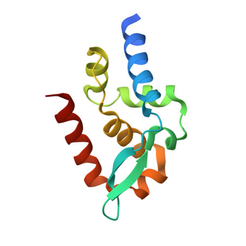
Structural comparison of the C-terminal domain of functionally divergent lyssavirus P proteins.

Sugiyama, A.Nomai, T.Jiang, X.Minami, M.Yao, M.Maenaka, K.Ito, N.Gooley, P.R.Moseley, G.W.Ose, T.

(2020) Biochem Biophys Res Commun 529: 507-512

  • DOI: https://doi.org/10.1016/j.bbrc.2020.05.195
  • Primary Citation of Related Structures:  
    7C20, 7C21

  • PubMed Abstract: 

    Lyssavirus P protein is a multifunctional protein that interacts with numerous host-cell proteins. The C-terminal domain (CTD) of P is important for inhibition of JAK-STAT signaling enabling the virus to evade host immunity. Several regions on the surface of rabies virus P are reported to interact with host factors. Among them, an extended, discrete hydrophobic patch of P CTD is notable. Although structures of P CTD of two strains of rabies virus, and of mokola virus have been solved, the structure of P CTD for Duvenhage virus, which is functionally divergent from these species for immune evasion function, is not known. Here, we analyze the structures of P CTD of Duvenhage and of a distinct rabies virus strain to gain further insight on the nature and potential function of the hydrophobic surface. Molecular contacts in crystals suggest that the hydrophobic patch is important to intermolecular interactions with other proteins, which differ between the lyssavirus species.


  • Organizational Affiliation

    Faculty of Advanced Life Science, Hokkaido University, Kita-10, Nishi-8, Kita-ku, Sapporo, 060-0810, Japan.


Macromolecules
Find similar proteins by:  (by identity cutoff)  |  3D Structure
Entity ID: 1
MoleculeChains Sequence LengthOrganismDetailsImage
Phosphoprotein112Lyssavirus duvenhageMutation(s): 0 
UniProt
Find proteins for O56774 (Duvenhage virus)
Explore O56774 
Go to UniProtKB:  O56774
Entity Groups  
Sequence Clusters30% Identity50% Identity70% Identity90% Identity95% Identity100% Identity
UniProt GroupO56774
Sequence Annotations
Expand
  • Reference Sequence
Experimental Data & Validation

Experimental Data

  • Method: X-RAY DIFFRACTION
  • Resolution: 1.95 Å
  • R-Value Free: 0.265 
  • R-Value Work: 0.198 
  • R-Value Observed: 0.203 
  • Space Group: C 1 2 1
Unit Cell:
Length ( Å )Angle ( ˚ )
a = 95.19α = 90
b = 49.52β = 99.75
c = 25.28γ = 90
Software Package:
Software NamePurpose
PHENIXrefinement
XDSdata reduction
XSCALEdata scaling
MOLREPphasing

Structure Validation

View Full Validation Report


Entry History & Funding Information

Deposition Data


Funding OrganizationLocationGrant Number
Japan Society for the Promotion of Science (JSPS)Japan17K07296

Revision History  (Full details and data files)

  • Version 1.0: 2021-03-17
    Type: Initial release
  • Version 1.1: 2023-11-29
    Changes: Data collection, Database references, Refinement description