7PGX | pdb_00007pgx

Structure of dark-adapted AsLOV2 wild type


Experimental Data Snapshot

  • Method: X-RAY DIFFRACTION
  • Resolution: 1.00 Å
  • R-Value Free: 
    0.147 (Depositor), 0.150 (DCC) 
  • R-Value Work: 
    0.114 (Depositor), 0.120 (DCC) 

Starting Model: experimental
View more details

wwPDB Validation   3D Report Full Report


Ligand Structure Quality Assessment 

Created with Raphaël 2.3.0Worse 01 BetterLigand structure goodness of fit to experimental dataBest fitted FMNClick on this verticalbar to view detailsBest fitted PEGClick on this verticalbar to view details

This is version 1.2 of the entry. See complete history


Literature

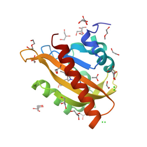
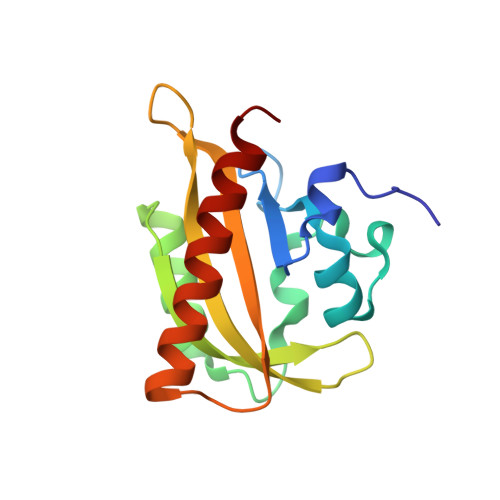
Signal transduction in light-oxygen-voltage receptors lacking the active-site glutamine.

Dietler, J.Gelfert, R.Kaiser, J.Borin, V.Renzl, C.Pilsl, S.Ranzani, A.T.Garcia de Fuentes, A.Gleichmann, T.Diensthuber, R.P.Weyand, M.Mayer, G.Schapiro, I.Moglich, A.

(2022) Nat Commun 13: 2618-2618

  • DOI: https://doi.org/10.1038/s41467-022-30252-4
  • Primary Citation of Related Structures:  
    7PGX, 7PGY, 7PGZ, 7PH0

  • PubMed Abstract: 

    In nature as in biotechnology, light-oxygen-voltage photoreceptors perceive blue light to elicit spatiotemporally defined cellular responses. Photon absorption drives thioadduct formation between a conserved cysteine and the flavin chromophore. An equally conserved, proximal glutamine processes the resultant flavin protonation into downstream hydrogen-bond rearrangements. Here, we report that this glutamine, long deemed essential, is generally dispensable. In its absence, several light-oxygen-voltage receptors invariably retained productive, if often attenuated, signaling responses. Structures of a light-oxygen-voltage paradigm at around 1 Å resolution revealed highly similar light-induced conformational changes, irrespective of whether the glutamine is present. Naturally occurring, glutamine-deficient light-oxygen-voltage receptors likely serve as bona fide photoreceptors, as we showcase for a diguanylate cyclase. We propose that without the glutamine, water molecules transiently approach the chromophore and thus propagate flavin protonation downstream. Signaling without glutamine appears intrinsic to light-oxygen-voltage receptors, which pertains to biotechnological applications and suggests evolutionary descendance from redox-active flavoproteins.


  • Organizational Affiliation

    Department of Biochemistry, University of Bayreuth, 95447, Bayreuth, Germany.


Macromolecules
Find similar proteins by:  (by identity cutoff)  |  3D Structure
Entity ID: 1
MoleculeChains Sequence LengthOrganismDetailsImage
NPH1-1A [auth AAA]146Avena sativaMutation(s): 0 
Gene Names: NPH1-1
EC: 2.7.11.1
UniProt
Find proteins for O49003 (Avena sativa)
Explore O49003 
Go to UniProtKB:  O49003
Entity Groups  
Sequence Clusters30% Identity50% Identity70% Identity90% Identity95% Identity100% Identity
UniProt GroupO49003
Sequence Annotations
Expand
  • Reference Sequence
Small Molecules
Ligands 7 Unique
IDChains Name / Formula / InChI Key2D Diagram3D Interactions
FMN (Subject of Investigation/LOI)
Query on FMN

Download Ideal Coordinates CCD File 
S [auth AAA]FLAVIN MONONUCLEOTIDE
C17 H21 N4 O9 P
FVTCRASFADXXNN-SCRDCRAPSA-N
PEG (Subject of Investigation/LOI)
Query on PEG

Download Ideal Coordinates CCD File 
N [auth AAA],
O [auth AAA]
DI(HYDROXYETHYL)ETHER
C4 H10 O3
MTHSVFCYNBDYFN-UHFFFAOYSA-N
GOL
Query on GOL

Download Ideal Coordinates CCD File 
D [auth AAA]
E [auth AAA]
F [auth AAA]
G [auth AAA]
H [auth AAA]
GLYCEROL
C3 H8 O3
PEDCQBHIVMGVHV-UHFFFAOYSA-N
EDO
Query on EDO

Download Ideal Coordinates CCD File 
P [auth AAA],
Q [auth AAA]
1,2-ETHANEDIOL
C2 H6 O2
LYCAIKOWRPUZTN-UHFFFAOYSA-N
ACT
Query on ACT

Download Ideal Coordinates CCD File 
R [auth AAA]ACETATE ION
C2 H3 O2
QTBSBXVTEAMEQO-UHFFFAOYSA-M
CA
Query on CA

Download Ideal Coordinates CCD File 
B [auth AAA]CALCIUM ION
Ca
BHPQYMZQTOCNFJ-UHFFFAOYSA-N
CL
Query on CL

Download Ideal Coordinates CCD File 
C [auth AAA]CHLORIDE ION
Cl
VEXZGXHMUGYJMC-UHFFFAOYSA-M
Experimental Data & Validation

Experimental Data

  • Method: X-RAY DIFFRACTION
  • Resolution: 1.00 Å
  • R-Value Free:  0.147 (Depositor), 0.150 (DCC) 
  • R-Value Work:  0.114 (Depositor), 0.120 (DCC) 
Space Group: P 21 21 21
Unit Cell:
Length ( Å )Angle ( ˚ )
a = 35.5α = 90
b = 56.42β = 90
c = 66.66γ = 90
Software Package:
Software NamePurpose
REFMACrefinement
XDSdata reduction
pointlessdata scaling
PHASERphasing

Structure Validation

View Full Validation Report


Ligand Structure Quality Assessment 

Created with Raphaël 2.3.0Worse 01 BetterLigand structure goodness of fit to experimental dataBest fitted FMNClick on this verticalbar to view detailsBest fitted PEGClick on this verticalbar to view details

Entry History & Funding Information

Deposition Data


Funding OrganizationLocationGrant Number
German Research Foundation (DFG)GermanyMO2192/8-1

Revision History  (Full details and data files)

  • Version 1.0: 2022-05-11
    Type: Initial release
  • Version 1.1: 2022-05-25
    Changes: Database references
  • Version 1.2: 2024-01-31
    Changes: Data collection, Derived calculations, Refinement description