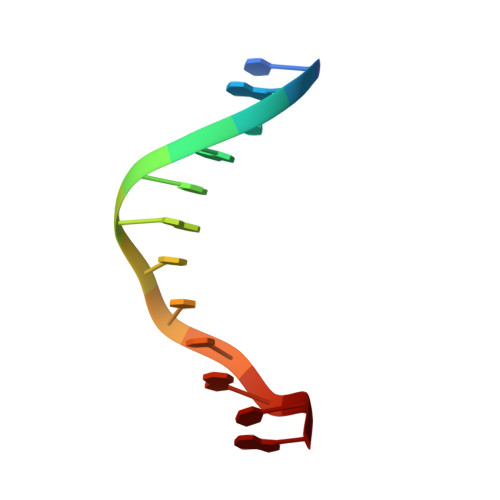
Binding of Hoechst 33258 to the minor groove of B-DNA.
Pjura, P.E., Grzeskowiak, K., Dickerson, R.E.(1987) J Mol Biol 197: 257-271
- PubMed: 2445998
- DOI: https://doi.org/10.1016/0022-2836(87)90123-9
- Primary Citation of Related Structures:
8BNA - PubMed Abstract:
An X-ray crystallographic structure analysis has been carried out on the complex between the antibiotic and DNA fluorochrome Hoechst 33258 and a synthetic B-DNA dodecamer of sequence C-G-C-G-A-A-T-T-C-G-C-G. The drug molecule, which can be schematized as: phenol-benzimidazole-benzimidazole-piperazine, sits within the minor groove in the A-T-T-C region of the DNA double helix, displacing the spine of hydration that is found in drug-free DNA. The NH groups of the benzimidazoles make bridging three-center hydrogen bonds between adenine N-3 and thymine O-2 atoms on the edges of base-pairs, in a manner both mimicking the spine of hydration and calling to mind the binding of the auti-tumor drug netropsin. Two conformers of Hoechst are seen in roughly equal populations, related by 180 degrees rotation about the central benzimidazole-benzimidazole bond: one form in which the piperazine ring extends out from the surface of the double helix, and another in which it is buried deep within the minor groove. Steric clash between the drug and DNA dictates that the phenol-benzimidazole-benzimidazole portion of Hoechst 33258 binds only to A.T regions of DNA, whereas the piperazine ring demands the wider groove characteristic of G.C regions. Hence, the piperazine ring suggests a possible G.C-reading element for synthetic DNA sequence-reading drug analogs.
Organizational Affiliation:
Molecular Biology Institute, University of California at Los Angeles 90024.