8R1V

Pseudomonas aeruginosa FabF C164A in complex with N-(1,5-dimethyl-3-oxo-2-phenyl-2,3-dihydro-1H-pyrazol-4-yl)-2-(4-methoxyphenoxy)acetamide


Experimental Data Snapshot

  • Method: X-RAY DIFFRACTION
  • Resolution: 2.09 Å
  • R-Value Free: 0.254 
  • R-Value Work: 0.189 

wwPDB Validation   3D Report Full Report


Ligand Structure Quality Assessment 


This is version 1.1 of the entry. See complete history


Literature

Design, quality and validation of the EU-OPENSCREEN fragment library poised to a high-throughput screening collection.

Jalencas, X.Berg, H.Espeland, L.O.Sreeramulu, S.Kinnen, F.Richter, C.Georgiou, C.Yadrykhinsky, V.Specker, E.Jaudzems, K.Miletic, T.Harmel, R.Gribbon, P.Schwalbe, H.Brenk, R.Jirgensons, A.Zaliani, A.Mestres, J.

(2024) RSC Med Chem 15: 1176-1188

  • DOI: https://doi.org/10.1039/d3md00724c
  • Primary Citation of Related Structures:  
    8PJ0, 8R0I, 8R1V

  • PubMed Abstract: 

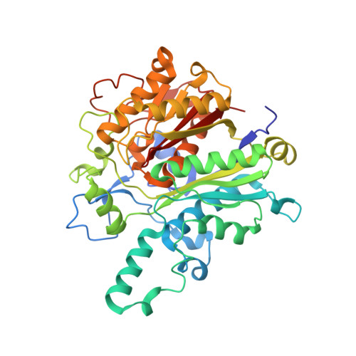
    The EU-OPENSCREEN (EU-OS) European Research Infrastructure Consortium (ERIC) is a multinational, not-for-profit initiative that integrates high-capacity screening platforms and chemistry groups across Europe to facilitate research in chemical biology and early drug discovery. Over the years, the EU-OS has assembled a high-throughput screening compound collection, the European Chemical Biology Library (ECBL), that contains approximately 100 000 commercially available small molecules and a growing number of thousands of academic compounds crowdsourced through our network of European and non-European chemists. As an extension of the ECBL, here we describe the computational design, quality control and use case screenings of the European Fragment Screening Library (EFSL) composed of 1056 mini and small chemical fragments selected from a substructure analysis of the ECBL. Access to the EFSL is open to researchers from both academia and industry. Using EFSL, eight fragment screening campaigns using different structural and biophysical methods have successfully identified fragment hits in the last two years. As one of the highlighted projects for antibiotics, we describe the screening by Bio-Layer Interferometry (BLI) of the EFSL, the identification of a 35 μM fragment hit targeting the beta-ketoacyl-ACP synthase 2 (FabF), its binding confirmation to the protein by X-ray crystallography (PDB 8PJ0), its subsequent rapid exploration of its surrounding chemical space through hit-picking of ECBL compounds that contain the fragment hit as a core substructure, and the final binding confirmation of two follow-up hits by X-ray crystallography (PDB 8R0I and 8R1V).


  • Organizational Affiliation

    Research Group on Systems Pharmacology, Research Program on Biomedical Informatics (GRIB), IMIM Hospital del Mar Medical Research Institute Parc de Recerca Biomèdica (PRBB), Doctor Aiguader 88 08003 Barcelona Spain jmestres@imim.es.


Macromolecules
Find similar proteins by:  (by identity cutoff)  |  3D Structure
Entity ID: 1
MoleculeChains Sequence LengthOrganismDetailsImage
3-oxoacyl-[acyl-carrier-protein] synthase 2
A, B
419Pseudomonas aeruginosaMutation(s): 1 
Gene Names: fabF1PA2965
EC: 2.3.1.179
UniProt
Find proteins for G3XDA2 (Pseudomonas aeruginosa (strain ATCC 15692 / DSM 22644 / CIP 104116 / JCM 14847 / LMG 12228 / 1C / PRS 101 / PAO1))
Explore G3XDA2 
Go to UniProtKB:  G3XDA2
Entity Groups  
Sequence Clusters30% Identity50% Identity70% Identity90% Identity95% Identity100% Identity
UniProt GroupG3XDA2
Sequence Annotations
Expand
  • Reference Sequence
Small Molecules
Ligands 3 Unique
IDChains Name / Formula / InChI Key2D Diagram3D Interactions
XJR (Subject of Investigation/LOI)
Query on XJR

Download Ideal Coordinates CCD File 
C [auth A],
H [auth B]
N-(1,5-dimethyl-3-oxo-2-phenyl-2,3-dihydro-1H-pyrazol-4-yl)-2-(4-methoxyphenoxy)acetamide
C20 H21 N3 O4
KFGTZDFVOVOTTI-UHFFFAOYSA-N
DMS
Query on DMS

Download Ideal Coordinates CCD File 
D [auth A],
I [auth B]
DIMETHYL SULFOXIDE
C2 H6 O S
IAZDPXIOMUYVGZ-UHFFFAOYSA-N
FMT
Query on FMT

Download Ideal Coordinates CCD File 
E [auth A]
F [auth A]
G [auth A]
J [auth B]
K [auth B]
E [auth A],
F [auth A],
G [auth A],
J [auth B],
K [auth B],
L [auth B],
M [auth B]
FORMIC ACID
C H2 O2
BDAGIHXWWSANSR-UHFFFAOYSA-N
Experimental Data & Validation

Experimental Data

  • Method: X-RAY DIFFRACTION
  • Resolution: 2.09 Å
  • R-Value Free: 0.254 
  • R-Value Work: 0.189 
  • Space Group: C 2 2 21
Unit Cell:
Length ( Å )Angle ( ˚ )
a = 100.947α = 90
b = 104.339β = 90
c = 144.117γ = 90
Software Package:
Software NamePurpose
REFMACrefinement
XDSdata reduction
Aimlessdata scaling
PHASERphasing

Structure Validation

View Full Validation Report


Ligand Structure Quality Assessment 


Entry History & Funding Information

Deposition Data


Funding OrganizationLocationGrant Number
Other governmentF-12602/4800005968
Research Council of NorwayNorway273588

Revision History  (Full details and data files)

  • Version 1.0: 2024-03-06
    Type: Initial release
  • Version 1.1: 2024-05-08
    Changes: Database references